Observed Characteristics:
Color:
- yellow
- purple-violet
- cinnamon-tan
- brown
Morphology:
- encrusting
- lobate
Consistency:
- soft
- crumbly
Locations:
- Colombia-San Andrés and Providencia Archipelago-Serrana Bank
- Colombia-San Andrés and Providencia Archipelago-San Andrés island
- Colombia-Santa Marta
- Colombia-Cartagena-Banco Salmedina
- Colombia-Islas del Rosario
- Colombia-Gulf of Urabá
- Panama-Bocas del Toro
- Puerto Rico-La Parguera
Species Description and Notes
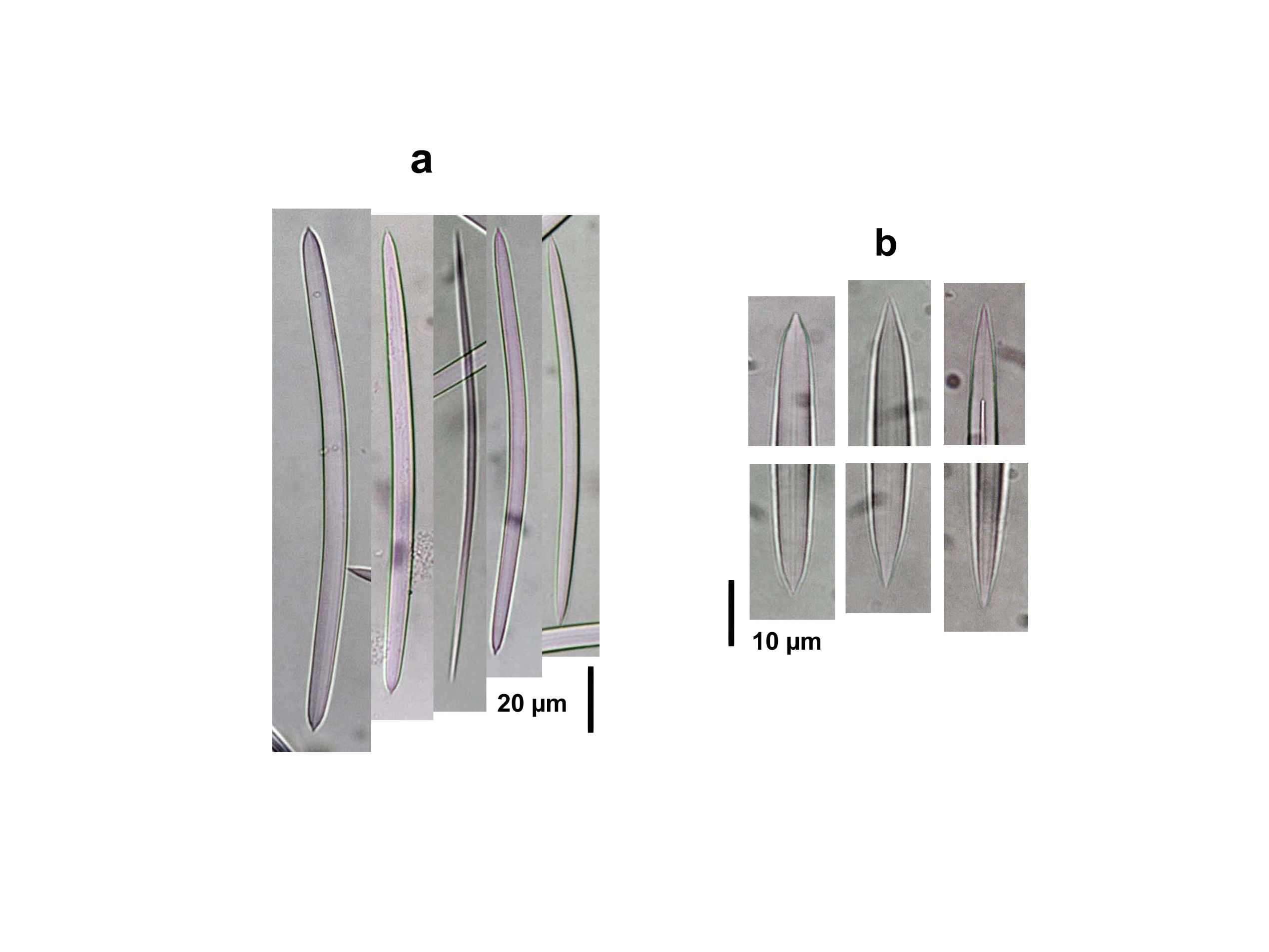
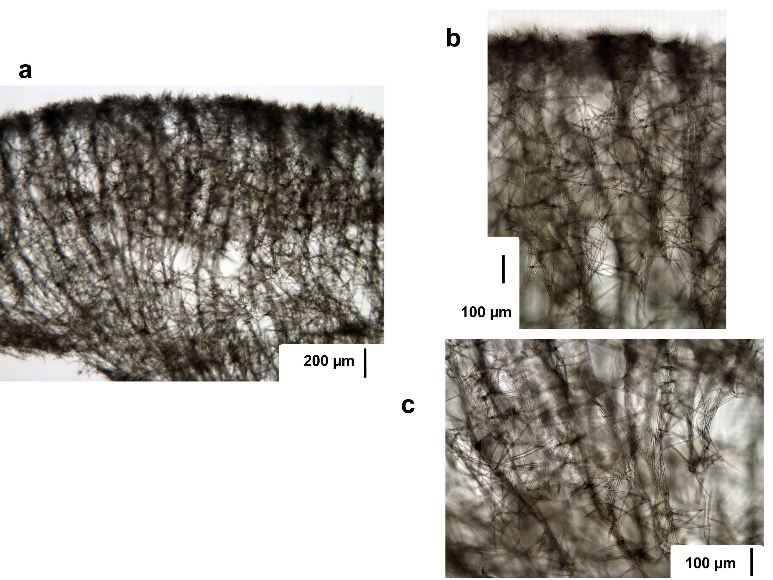
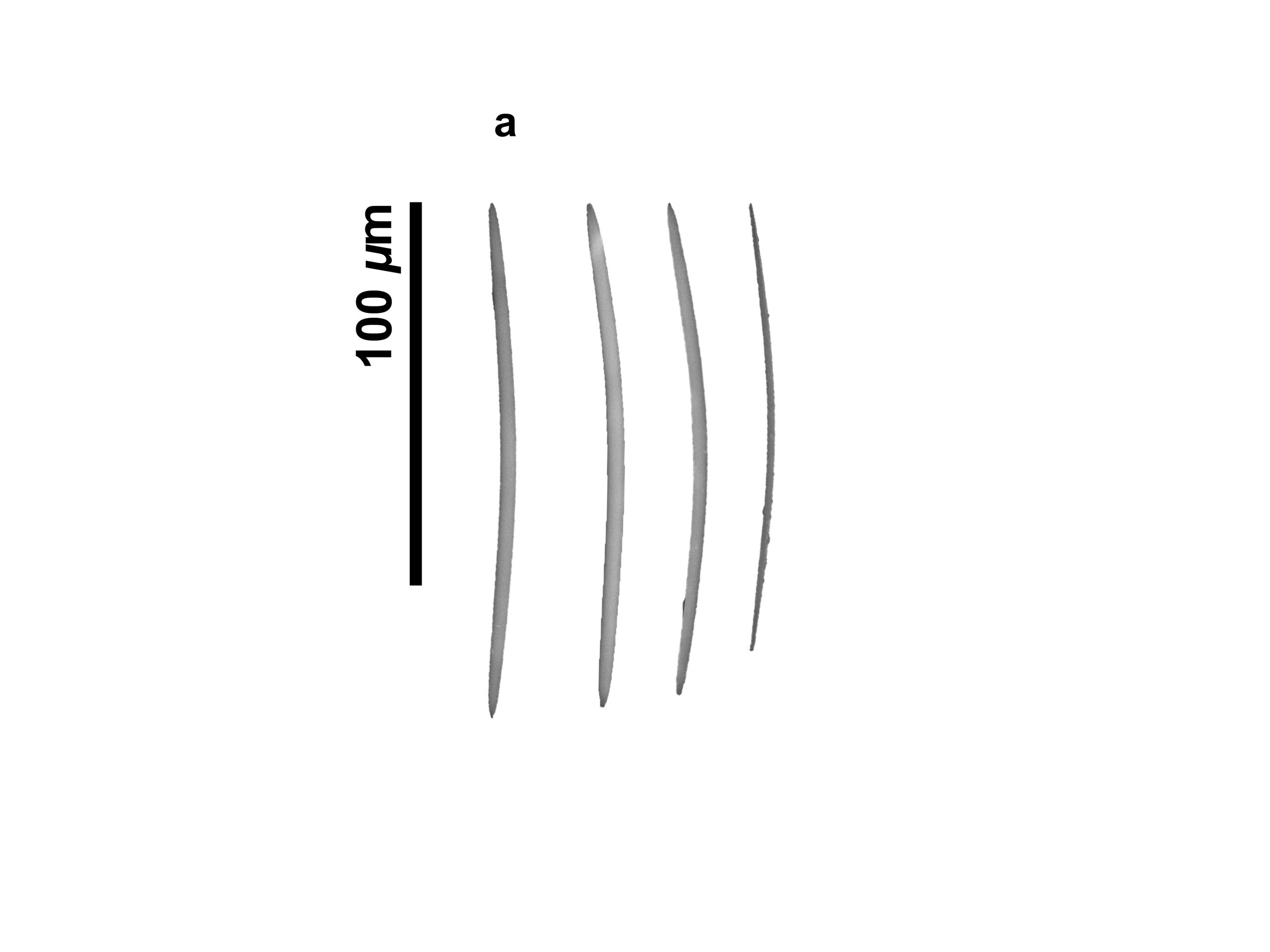
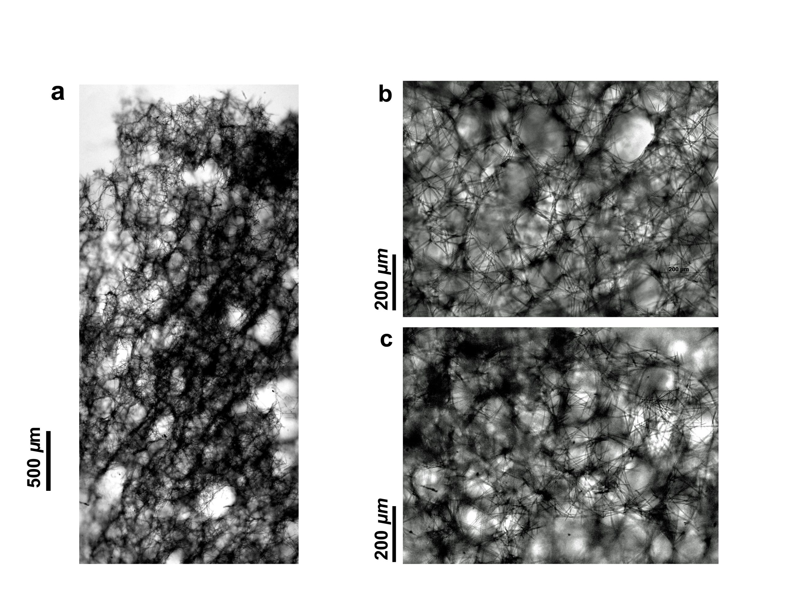
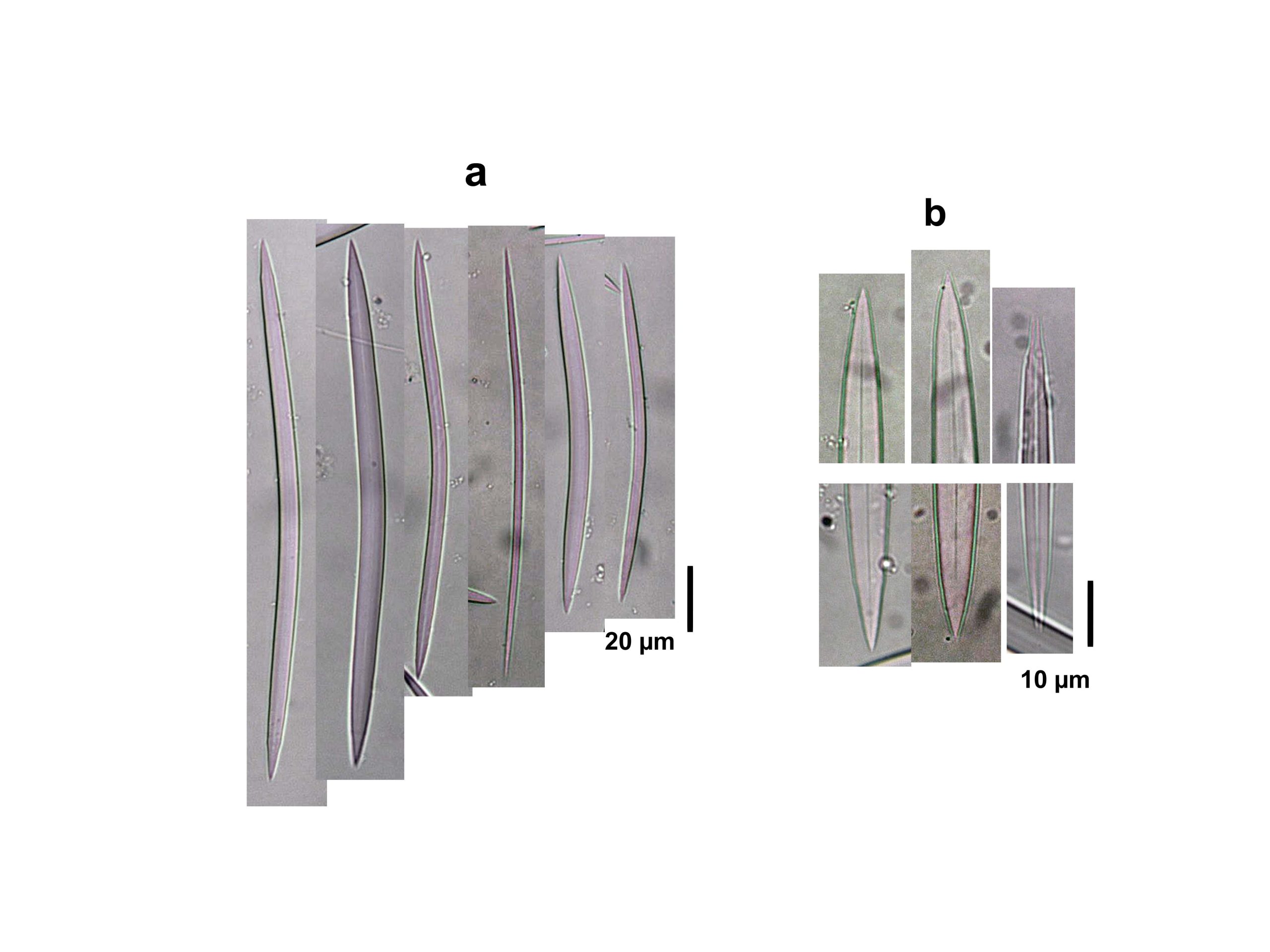
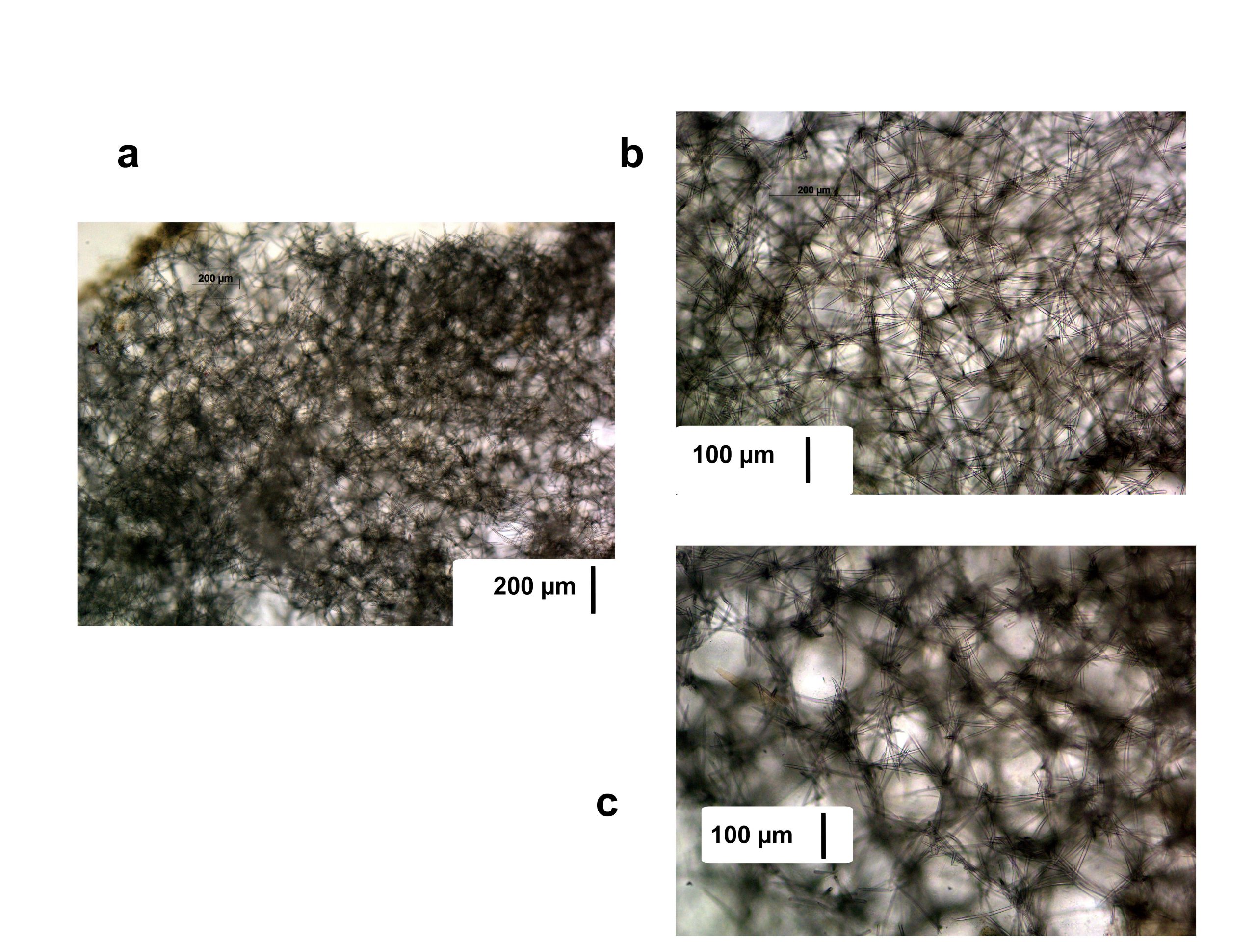
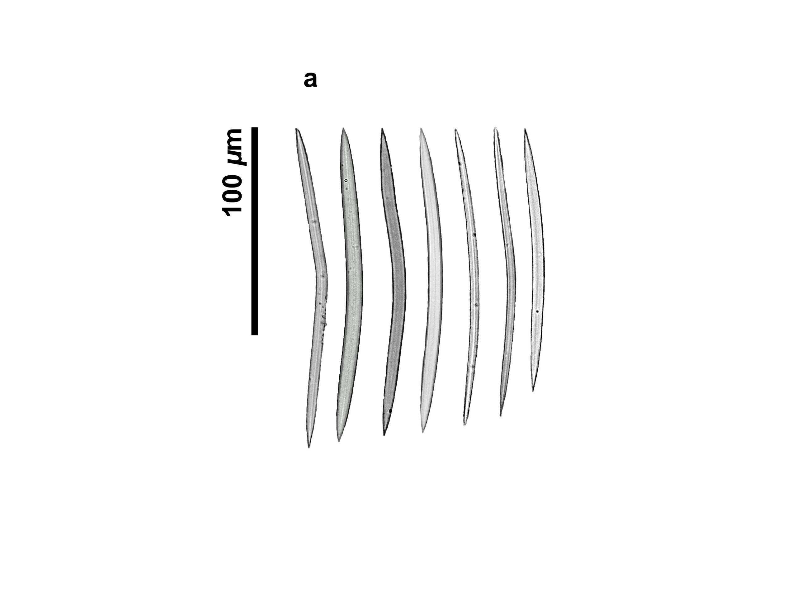
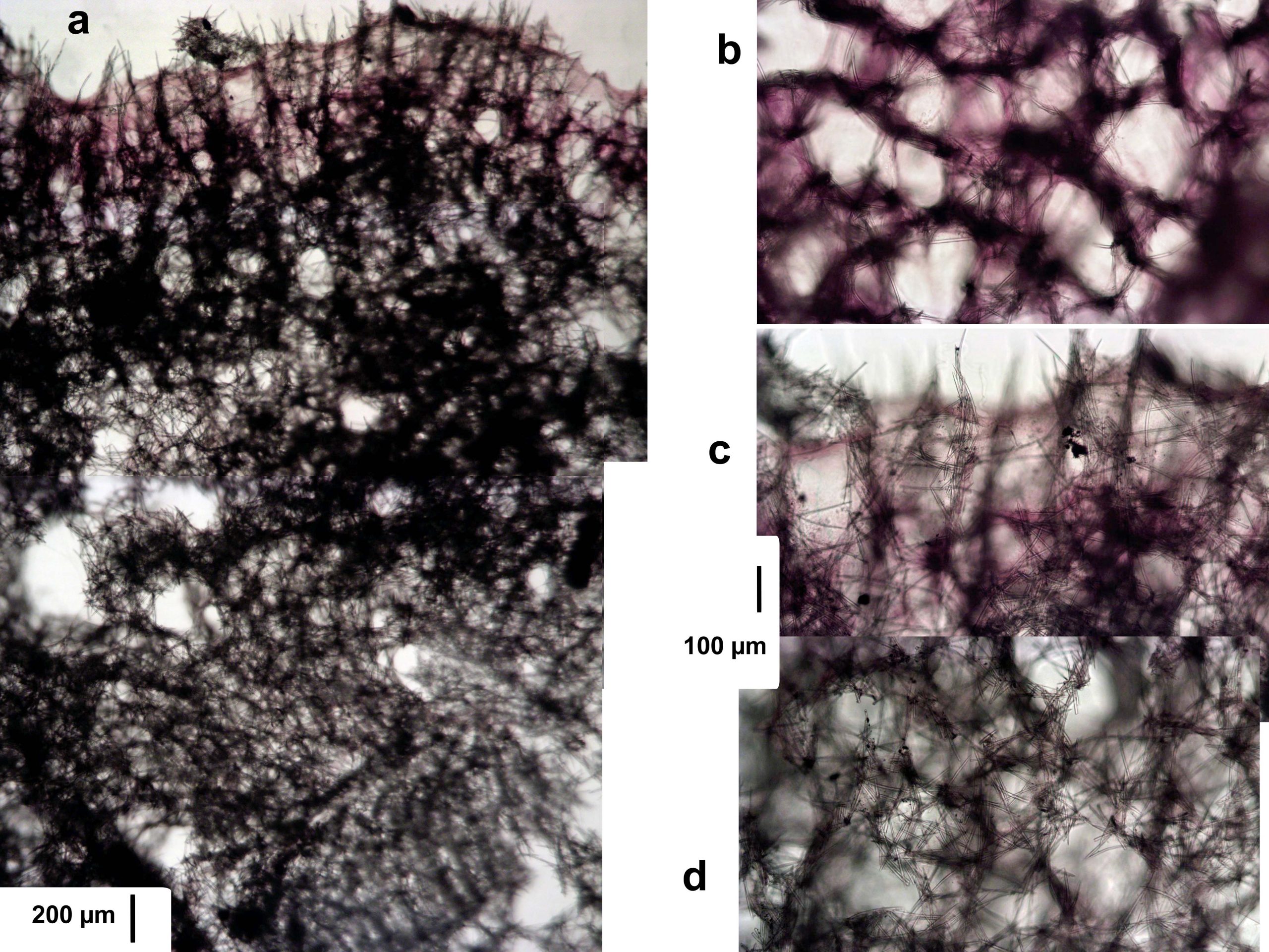
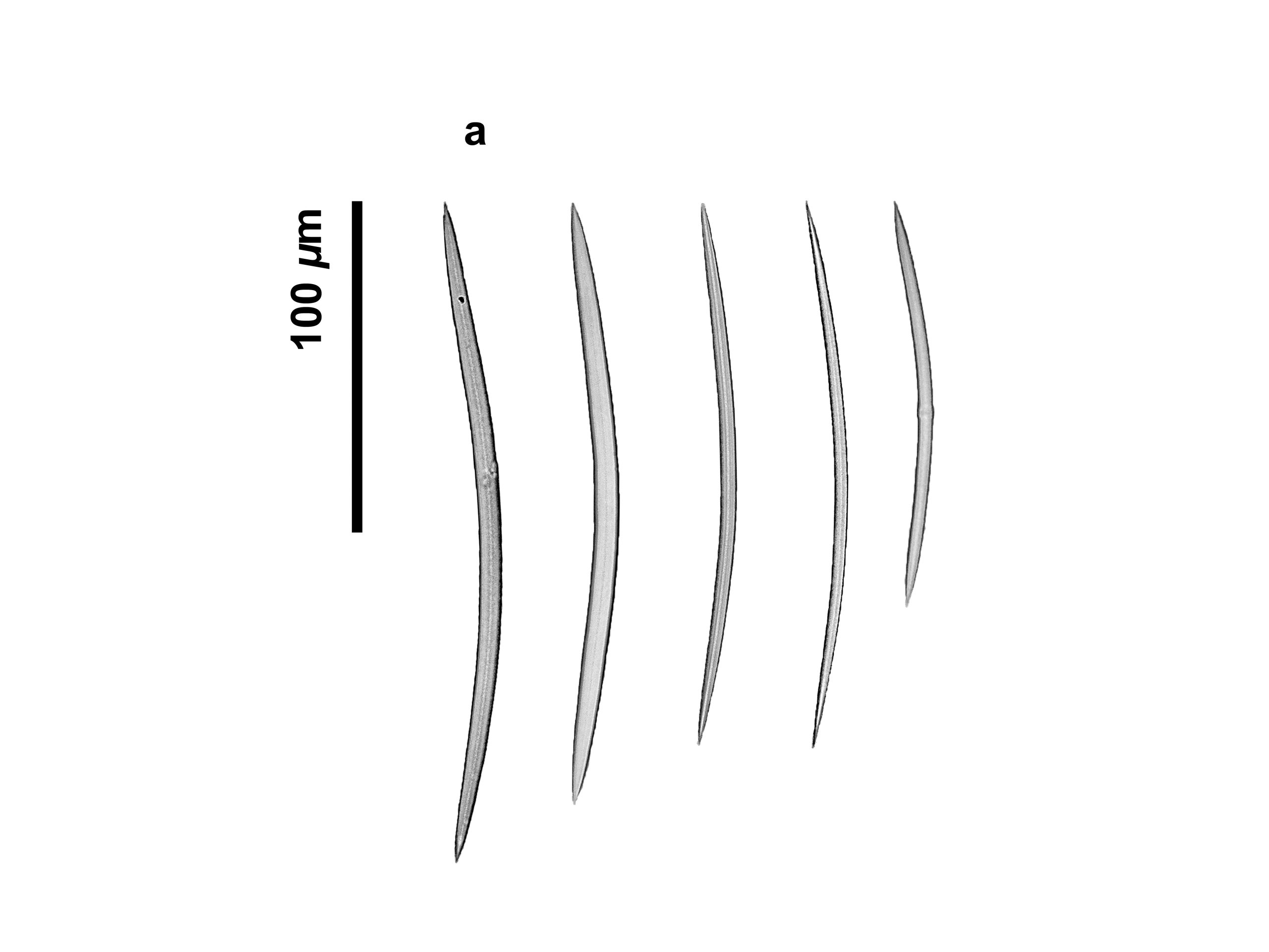
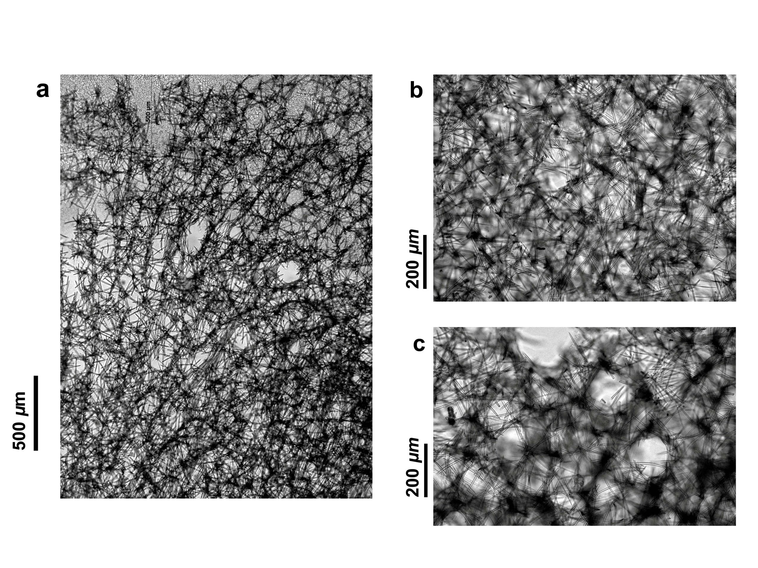
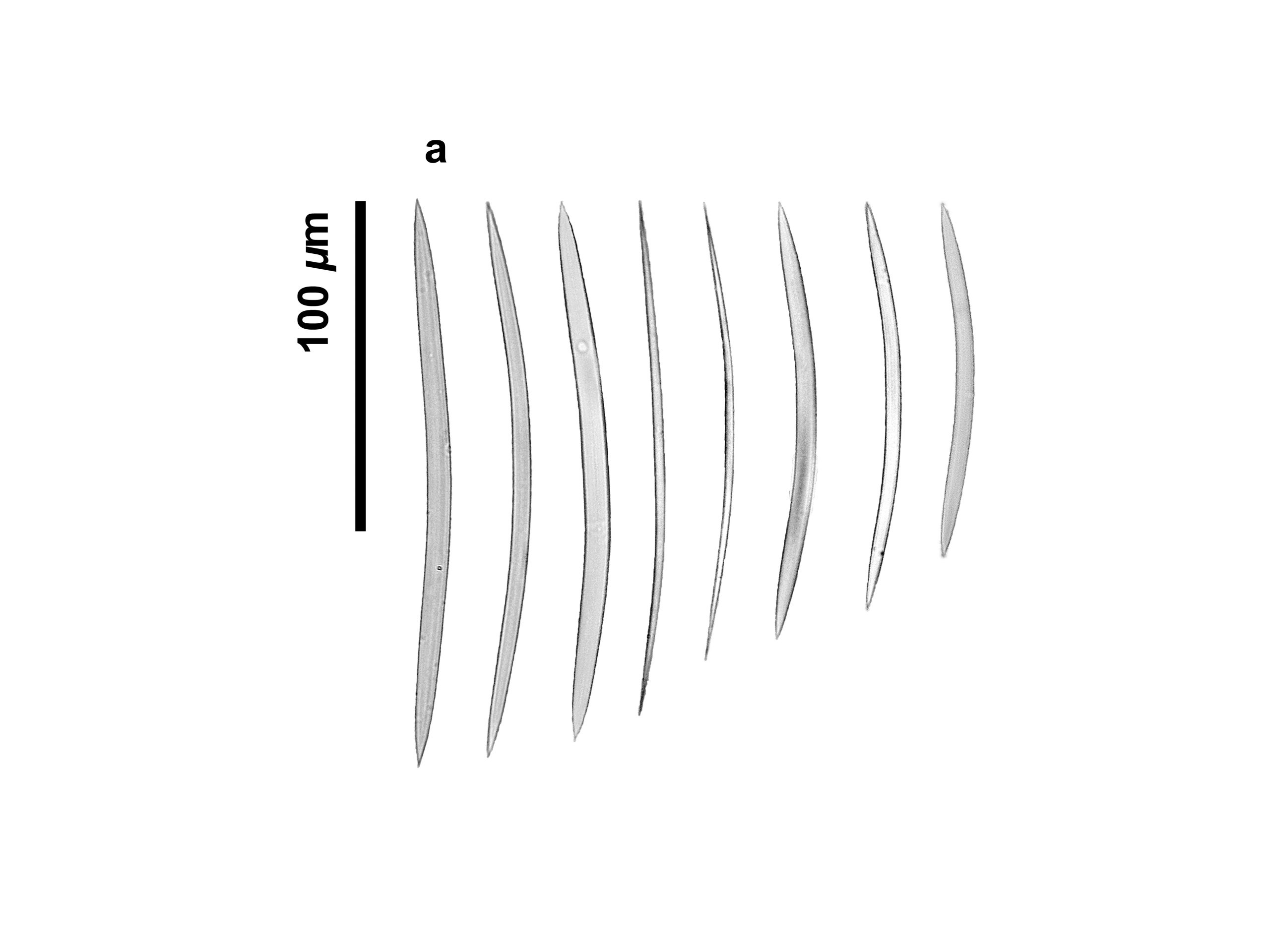
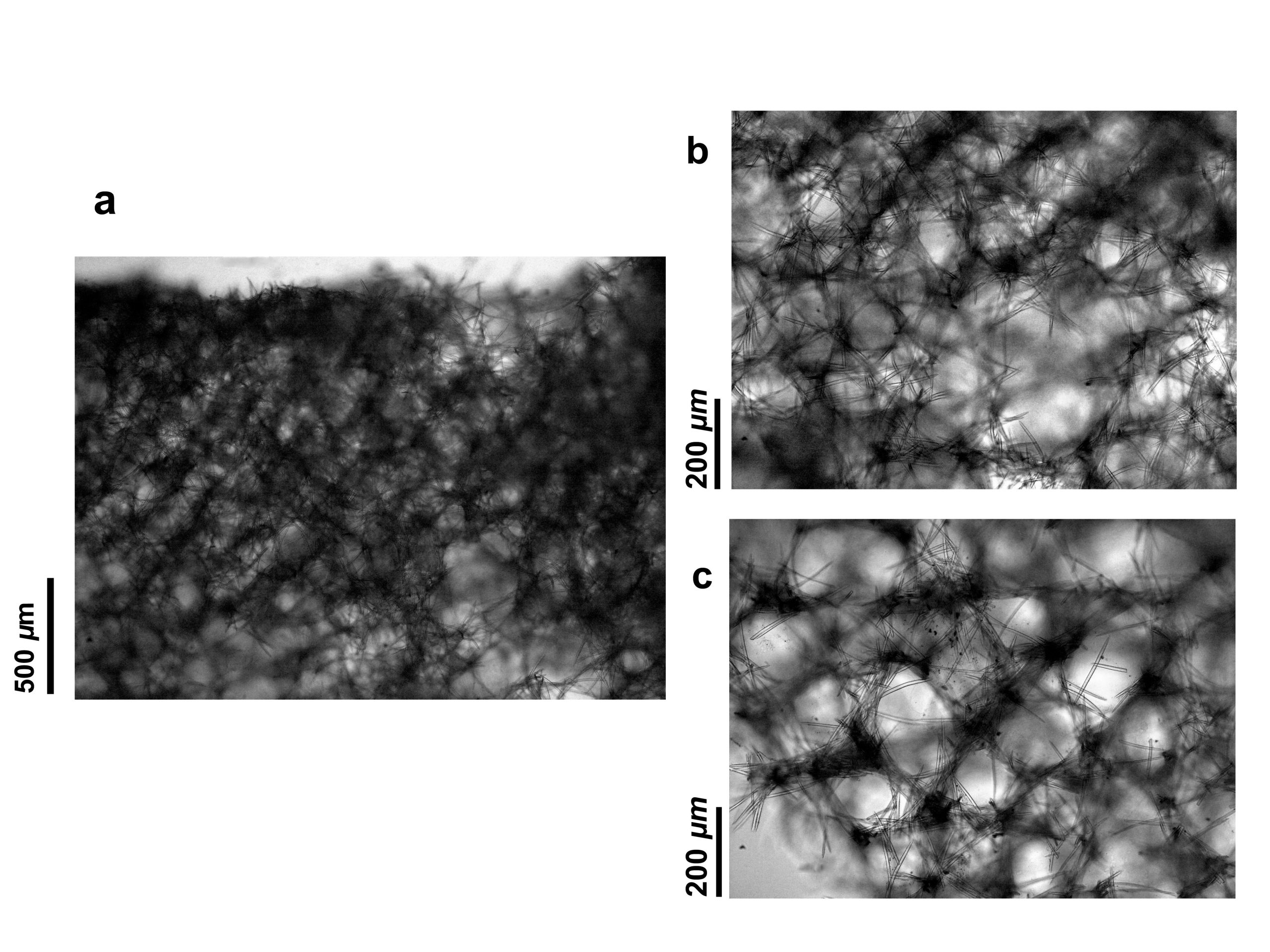
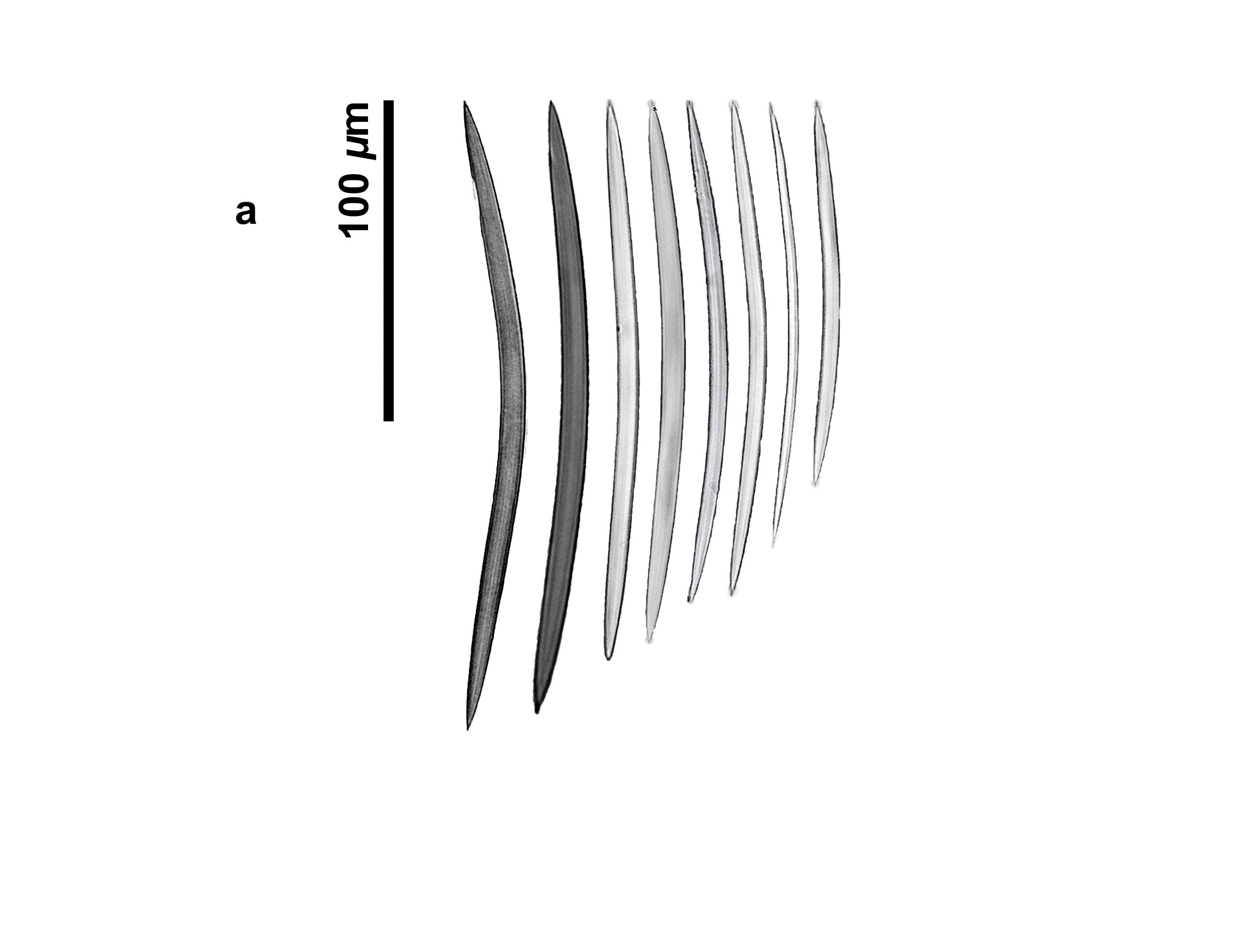
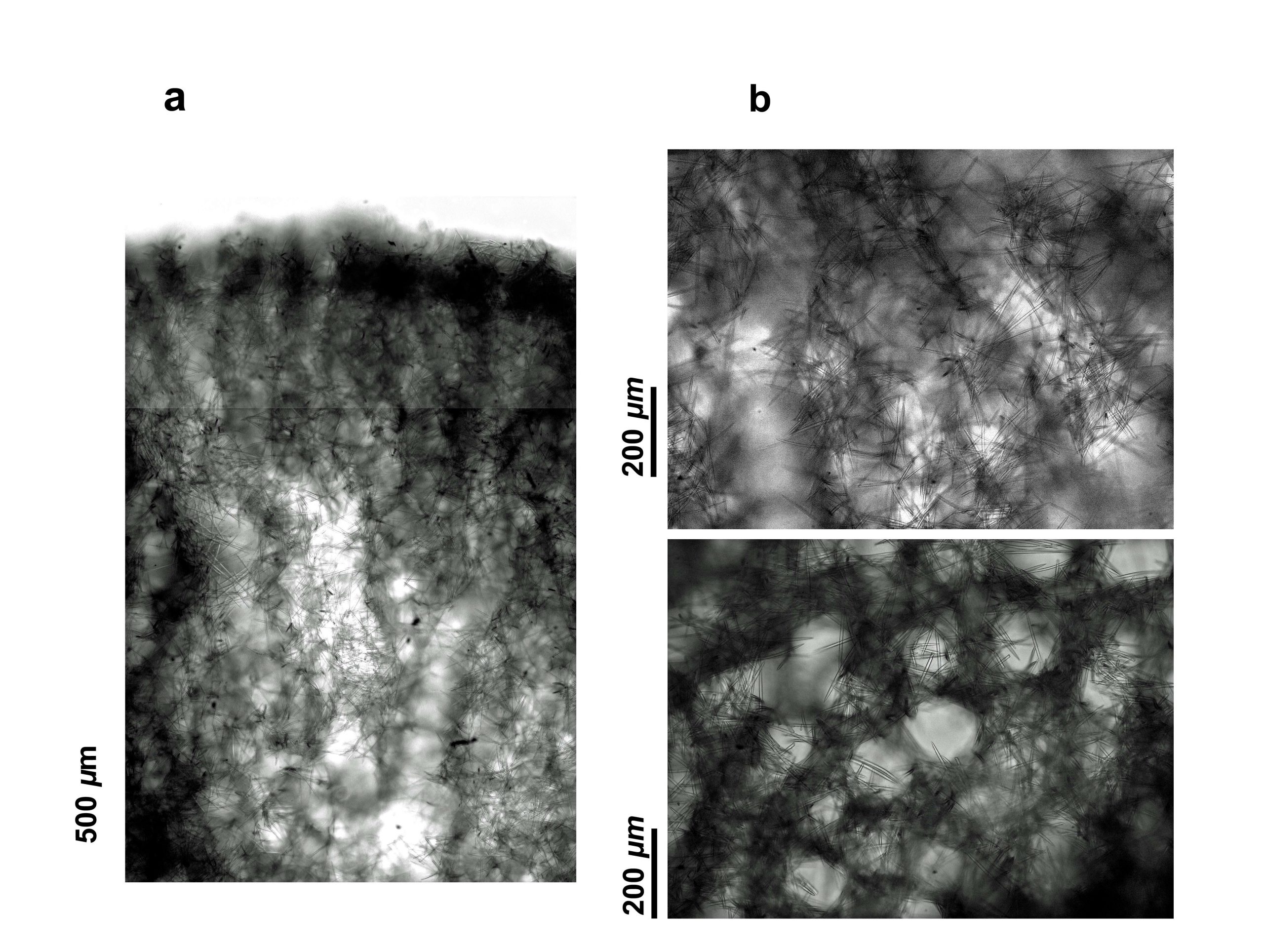
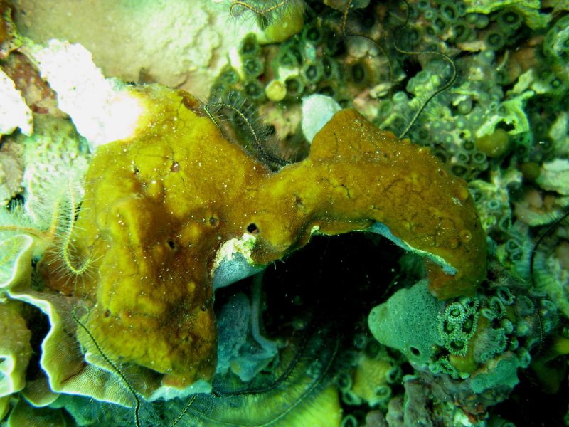
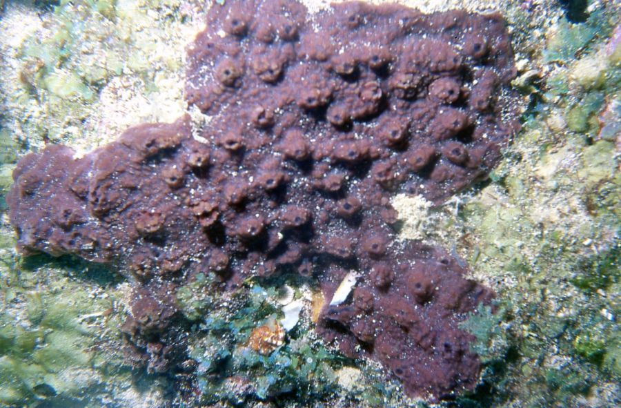
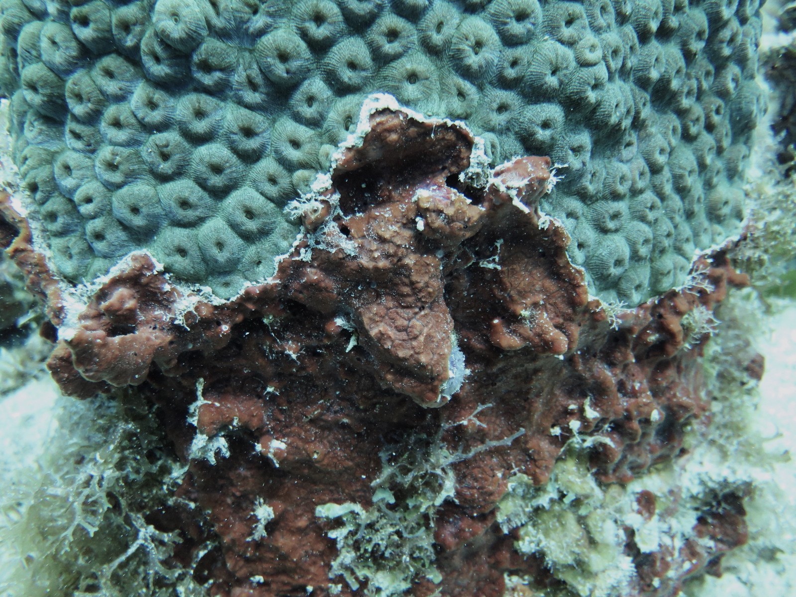
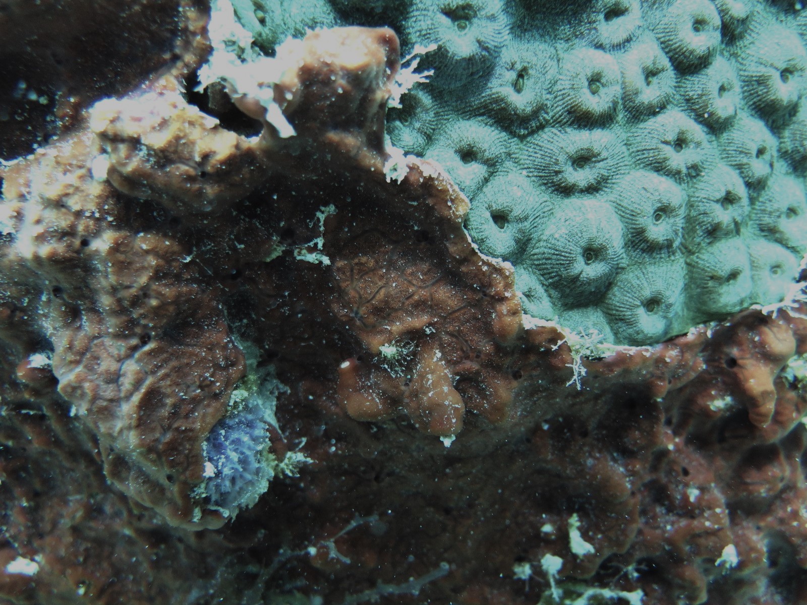
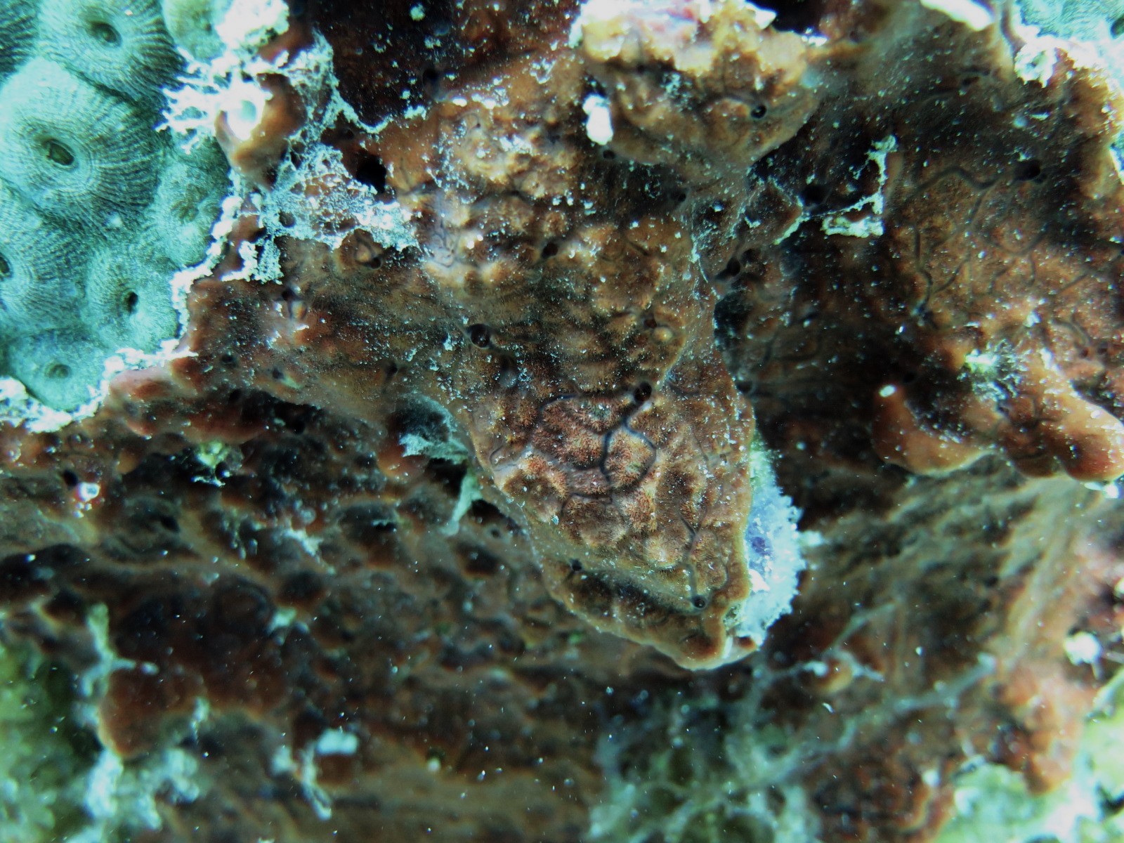
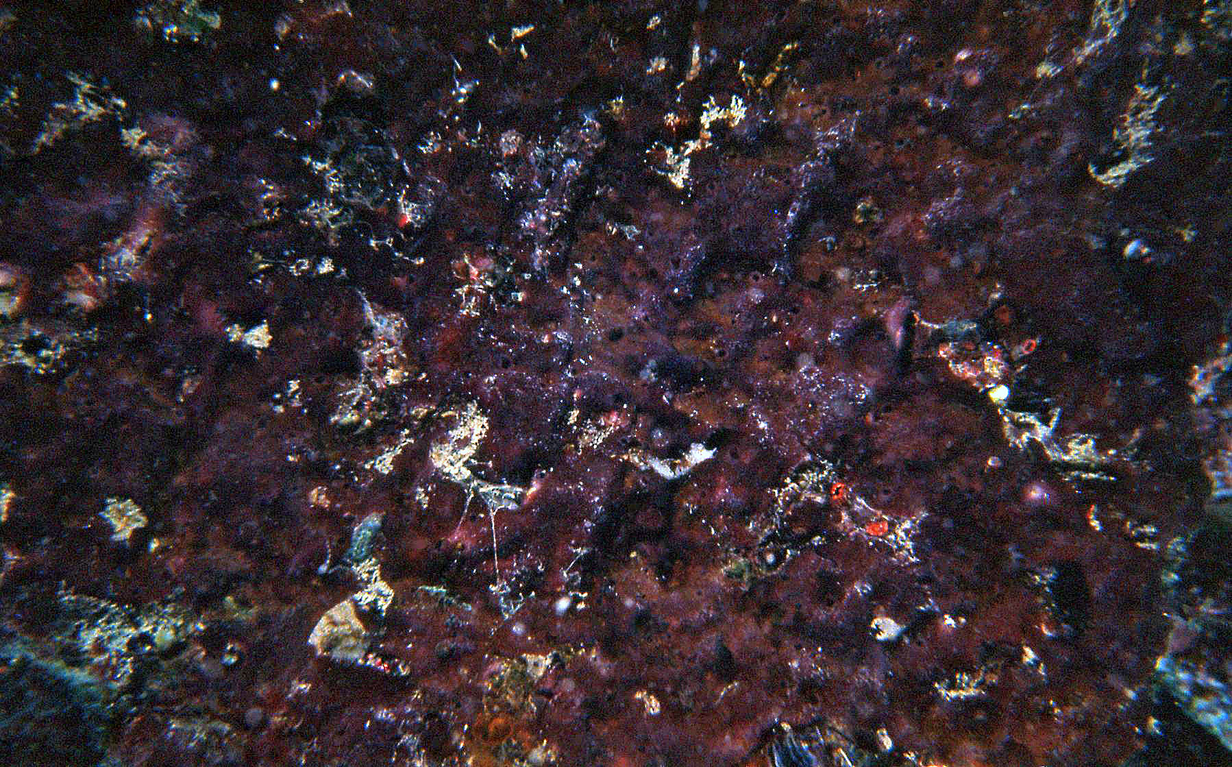
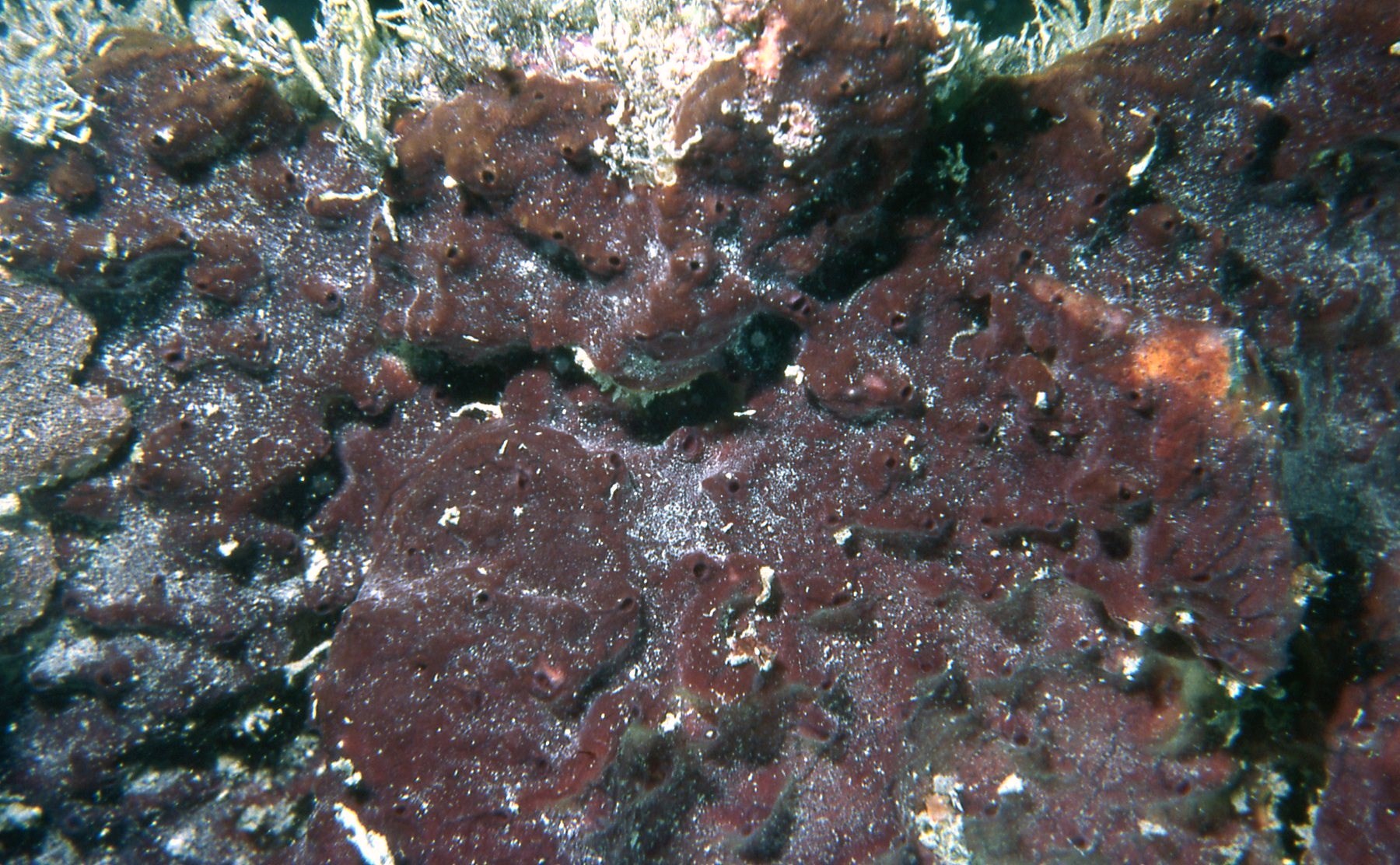
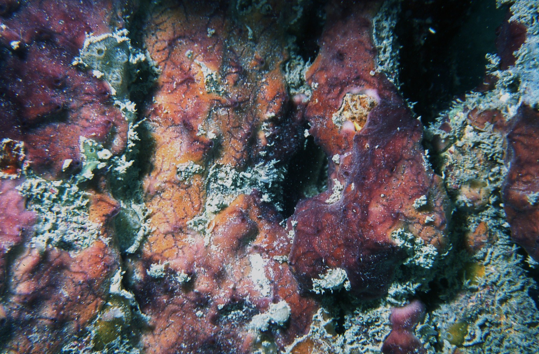
Description: Thin to thick encrustations, up to about 1 cm in thickness, covering several tens of cm of the substratum, which is often dead coral. The color can be burnt yellow to cinnamon-tan with purple tinges, or completely purplish brown, with the interior cream. The surface can be even or bumpy, with a network of slightly depressed veins which are subsurface canals, either converging in oscules or forming an irregular network. Oscules are dispersed, even or on top of mounds, up to about 4-5 mm in diameter, often with a membranous collar. Consistency is crumbly, easily tattered when handled; thinly encrusting specimens are generally soft. The skeleton is an anisotropic reticulation of irregularly ascending and diverging spicule tracts, 2-10 or more spicules in cross-section, 10-50 µm thick, separated some 50-130 µm, irregularly interconnected by single spicules or by spicule fascicles (sheets). Tracts end at the surface as spicule brushes; these, together with paratangentially placed, interconnecting spicules form a dense, confused ectosome, some 300-400 µm thick. Spicules are hastate oxea, stout, slightly curved, with thinner developmental stages, and ends short, acute, concave-conical, often slightly telescopic, 86-198 µm long by 2.0-10.5 µm thick. Sizes vary with locality, being larger in the continental coast of South America (e.g., mean 171.4 by 7.4 µm in the Gulf of Urabá), and shorter and thinner in far off oceanic locations (e.g., mean 130.1 by 3.8 µm in the San Andrés and Providencia Archipelago). For details in sizes see Vicente et al. (2019).
Notes: This species inhabits shallow to deep reefs, living exposed, often covering dead foliose or massive corals. In the third edition of the guide we called this species Neopetrosia sp.-“soft”. It may be initially confused in the field with N. proxima (Duchassaing & Michelotti, 1864), also pictured here, but can be distinguished by the usually softer texture and the lattice of canals visible at the surface. However, some of our specimes were hard enough for an initiall confusion, which was solved by looking at the tissue sections, which are more delicate in N. dendrocreavacea. I may also be confused in the field with Svenzea cristinae Alvarez, van Soest & Rützler, 2002, also pictured here, which is also thickly encrusting and soft, yellow to vinaceous, but has longer styles instead of oxea as spicules.
Author Reference: Vicente, Ríos, Zea & Toonen, 2019
Link: World Porifera Database
Tissue and Spicule Images
Images
dendrocrevacea

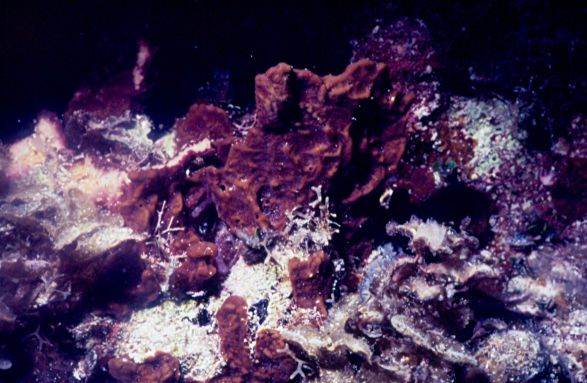
- Location: Panama-Bocas del Toro
- Photographer: Sven Zea
- Location: Panama-Bocas del Toro
- Photographer: Sven Zea
- Location: Panama-Bocas del Toro
- Photographer: Sven Zea
- Location: Puerto Rico-La Parguera
- Photographer: Sven Zea
- Location: Colombia-San Andrés and Providencia Archipelago-Serrana Bank
- Photographer: Sven Zea
- Location: Colombia-San Andrés and Providencia Archipelago-San Andrés island
- Photographer: Sven Zea
- Location: Colombia-San Andrés and Providencia Archipelago-San Andrés island
- Photographer: Sven Zea
- Location: Colombia-San Andrés and Providencia Archipelago-San Andrés island
- Photographer: Sven Zea
- Location: Colombia-Santa Marta
- Photographer: Sven Zea
- Location: Colombia-Santa Marta
- Photographer: Sven Zea
- Location: Colombia-Santa Marta
- Photographer: Sven Zea