Observed Characteristics:
Color:
- purple-violet
Morphology:
- massive
- branching
Consistency:
- hard
Locations:
- Colombia-Islas del Rosario
Species Description and Notes
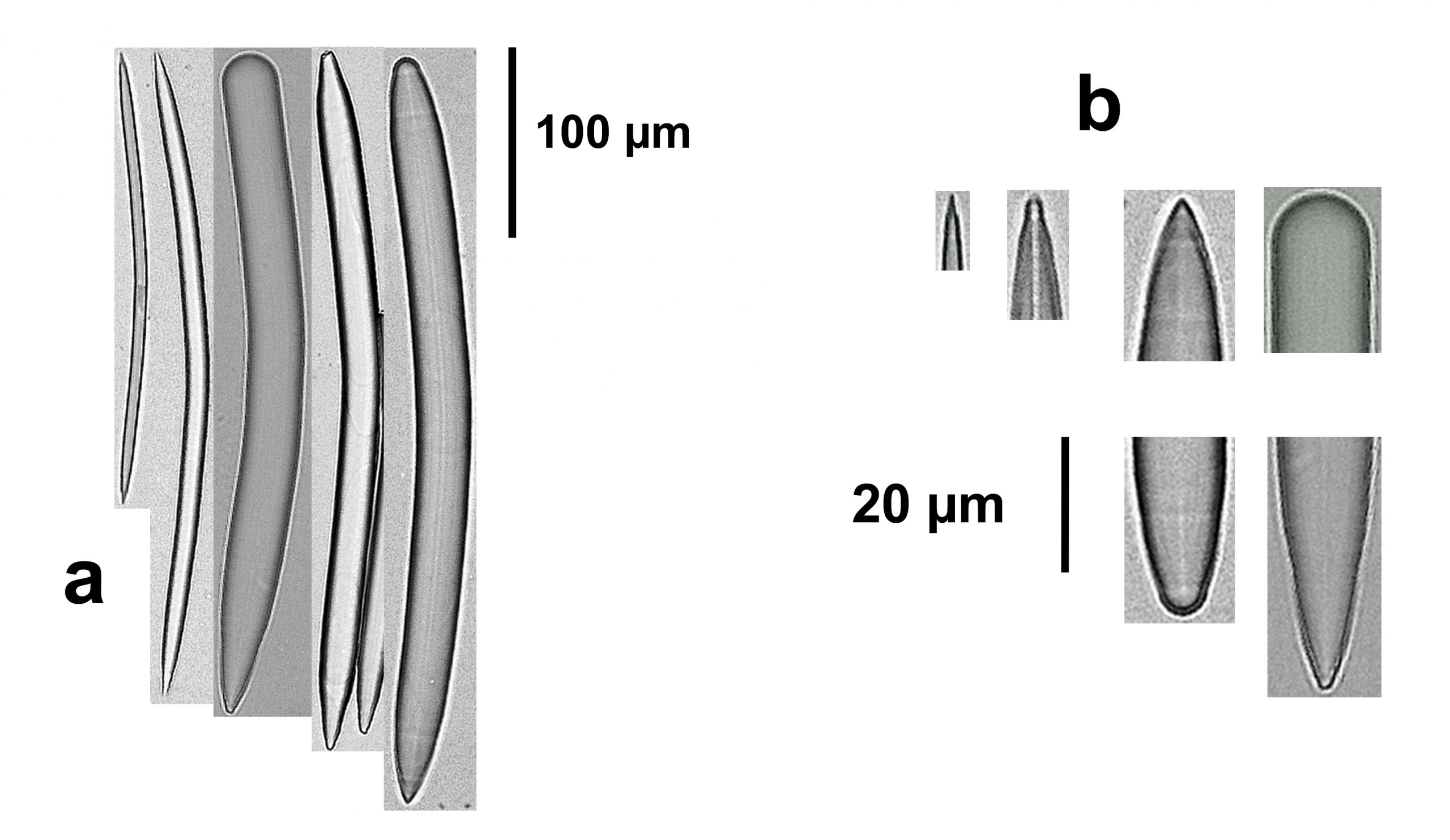
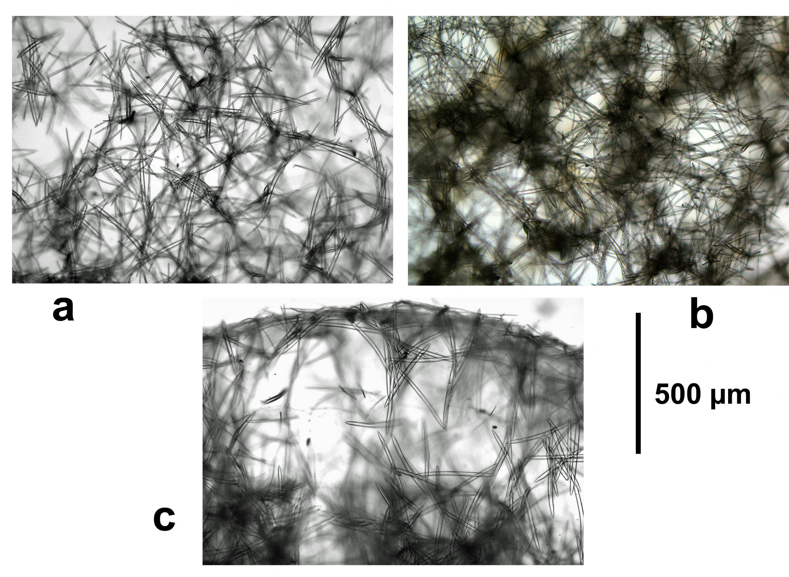
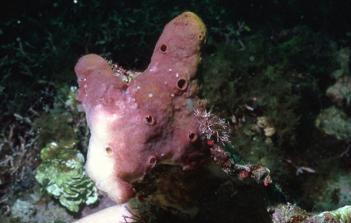
Description: Massive, with finger-like projections as primordial branches; size about 5 cm wide by 8-10 cm high. Surface even, micro rugose. Oscules relatively large, dispersed, 1-4 mm in diameter, with a slightly elevated edge. Color milky purple, with basal parts cream. Consistency hard, brittle. The ectosomal skeleton comprises an upper tangential reticulum of oxeas I (thinner, fusiform), somewhat confused, paucispicular (1–4 spicule tracts), forming meshes 60–150 μm in diameter, supported by a subsuperficial reticulum of thicker tracts of oxeas II (hastate, 3–7 spicules, 30–120 μm thick), with meshes 200–500 μm in diameter. The choanosomal skeleton is an isodictyal, rather confused reticulation, although ascending and interconnecting tracts can be discerned, 8–14 spicules and 240–260 μm thick, forming meshes of 300–700 μm in diameter, built by both types of oxeas. Near the sponge surface, growing layers are clearly distinct, but the skeleton becomes denser and confused as one goes deeper. The tracts have spongin cementing the spicules. Spicules are: oxeas in two size classes, (1) oxeas I, fusiform, slightly curved, with sharp points, 192-326 by 9.5-18.7 μm; (2) oxeas II, hastate, larger and thicker, with endings slightly blunt and sometimes mammiform or telescopic, or asymmetric turning into strongyloxeas and styles, 347-503 by 13-33.2 μm (description modified from Silva & Zea, 2017).
Notes: This is a shallow reef species. Upon collection, the specimen was confused with a branching Neopetrosia proxima (Duchassaing and Michelotti, 1864), also pictured here, but upon spicule examination it became clear, from the larger spicule size, that it was a species of Xestospongia. Once described, it was possible to assign the proper species name. The original material from Belize, found in cryptic habitats (1-18 m in depth), was encrusting to cushion shaped, with a knobbed surface and dispersed oscules, purple in color on top, with cream sides and base (Rützler et al., 2014). In addition to having the same two size classes of oxea, the studied specimen agrees with the Belizean original ones in color; its branch-like protuberances could be equivalent to the knobs of the Belizean specimens but with a greater growth. Interestingly, and to add to the variability of the species, in the studied specimen the first layer of the ectosome is built up exclusively of the thinner, smaller oxea, possibly as a new layer of growth. From its shape, color, and the two spicule size classes, this species is clearly distinct from the other Xestospongia species in the Caribbean.
Author Reference: Rützler, Piantoni, van Soest and Díaz, 2014
Link: World Porifera Database
Tissue and Spicule Images
Images
purpurea
- Location: Colombia-Islas del Rosario
- Photographer: Sven Zea
Observed Characteristics:
Color:
- yellow
Morphology:
- papillated
Consistency:
- soft
Locations:
- Colombia
- Panama
Species Description and Notes
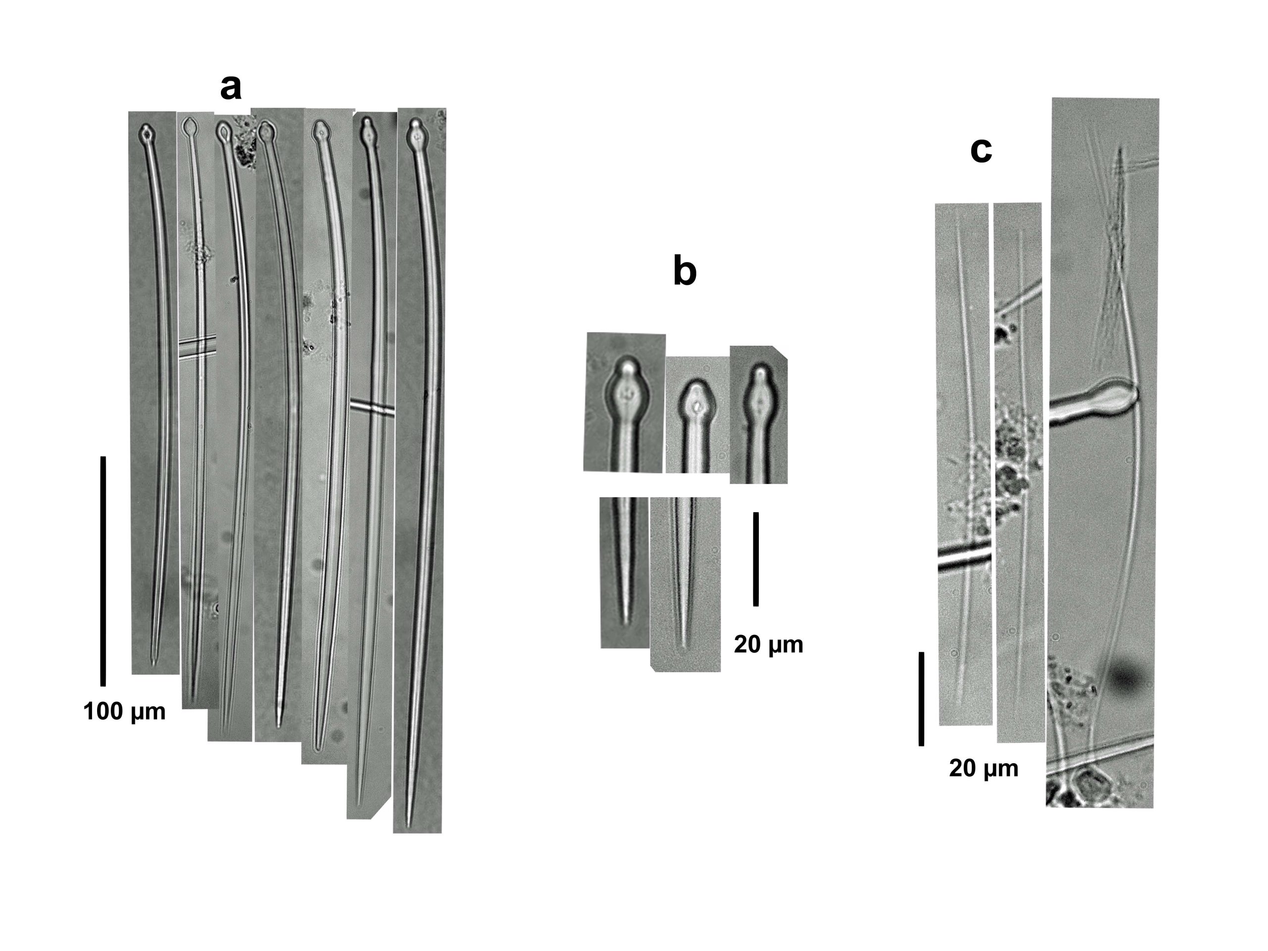
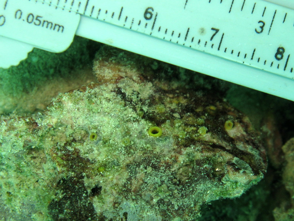
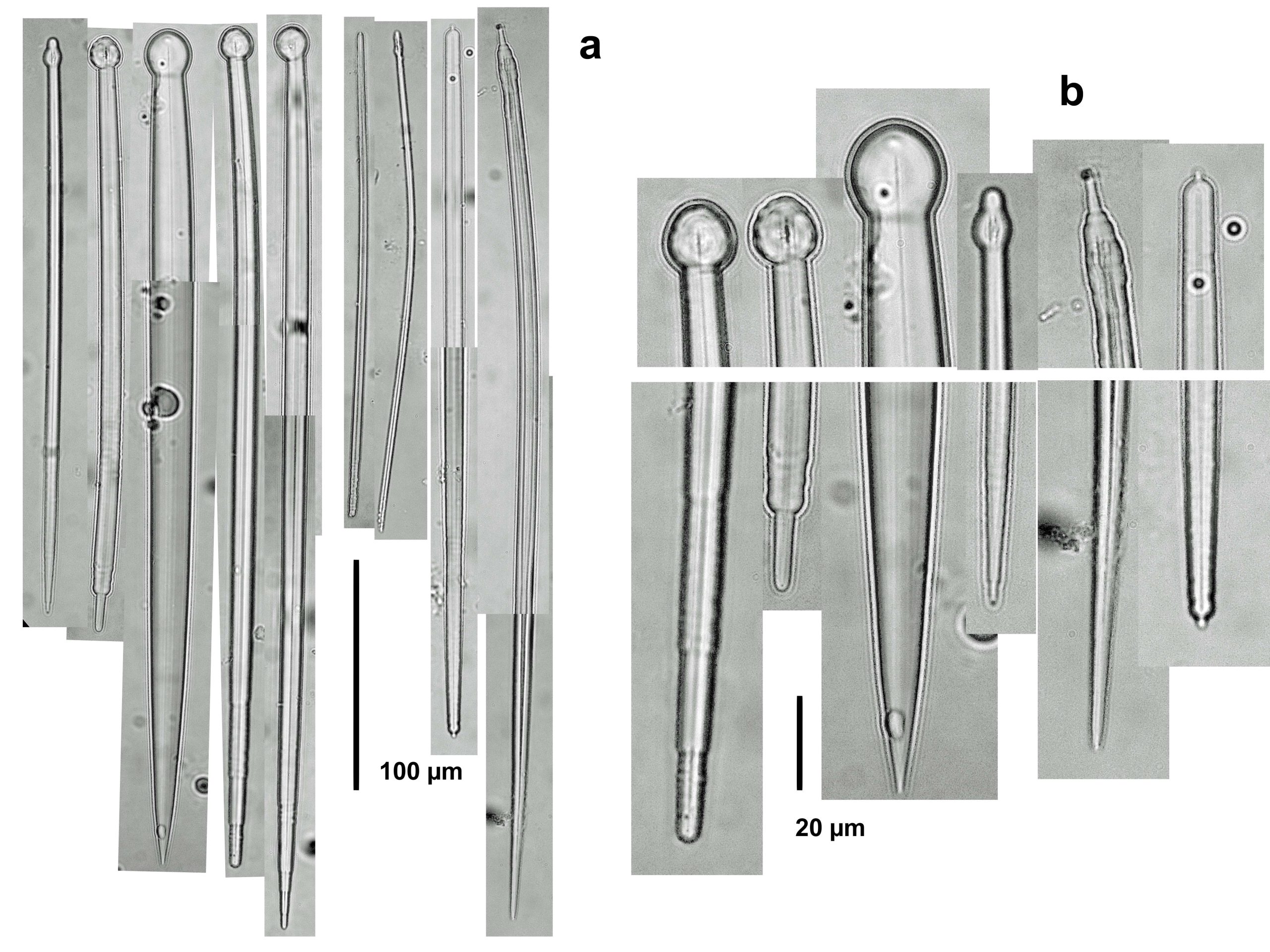
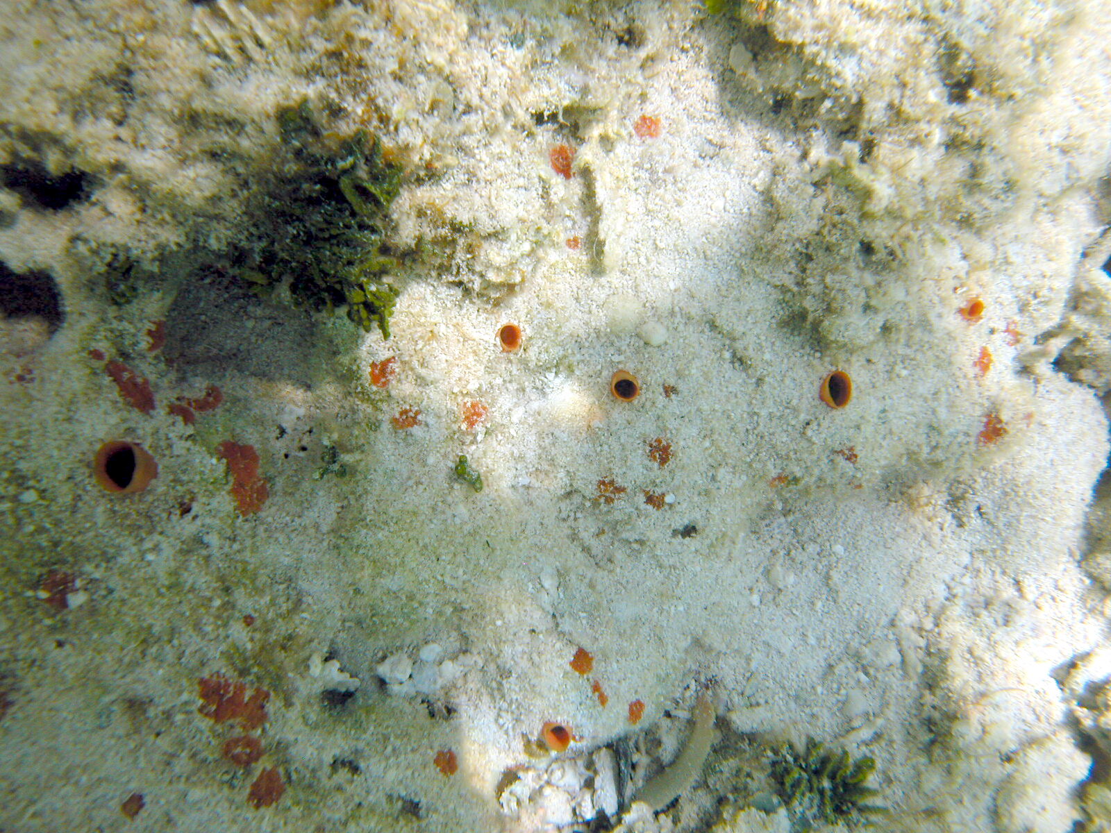
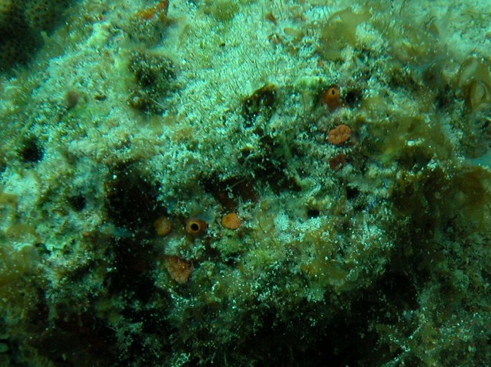
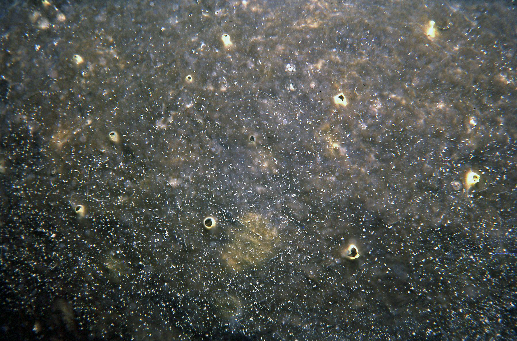
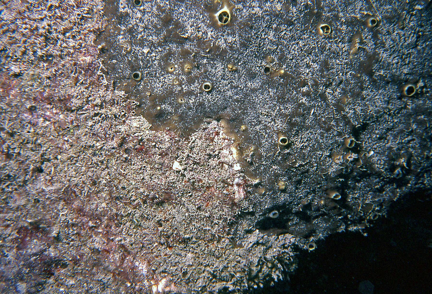
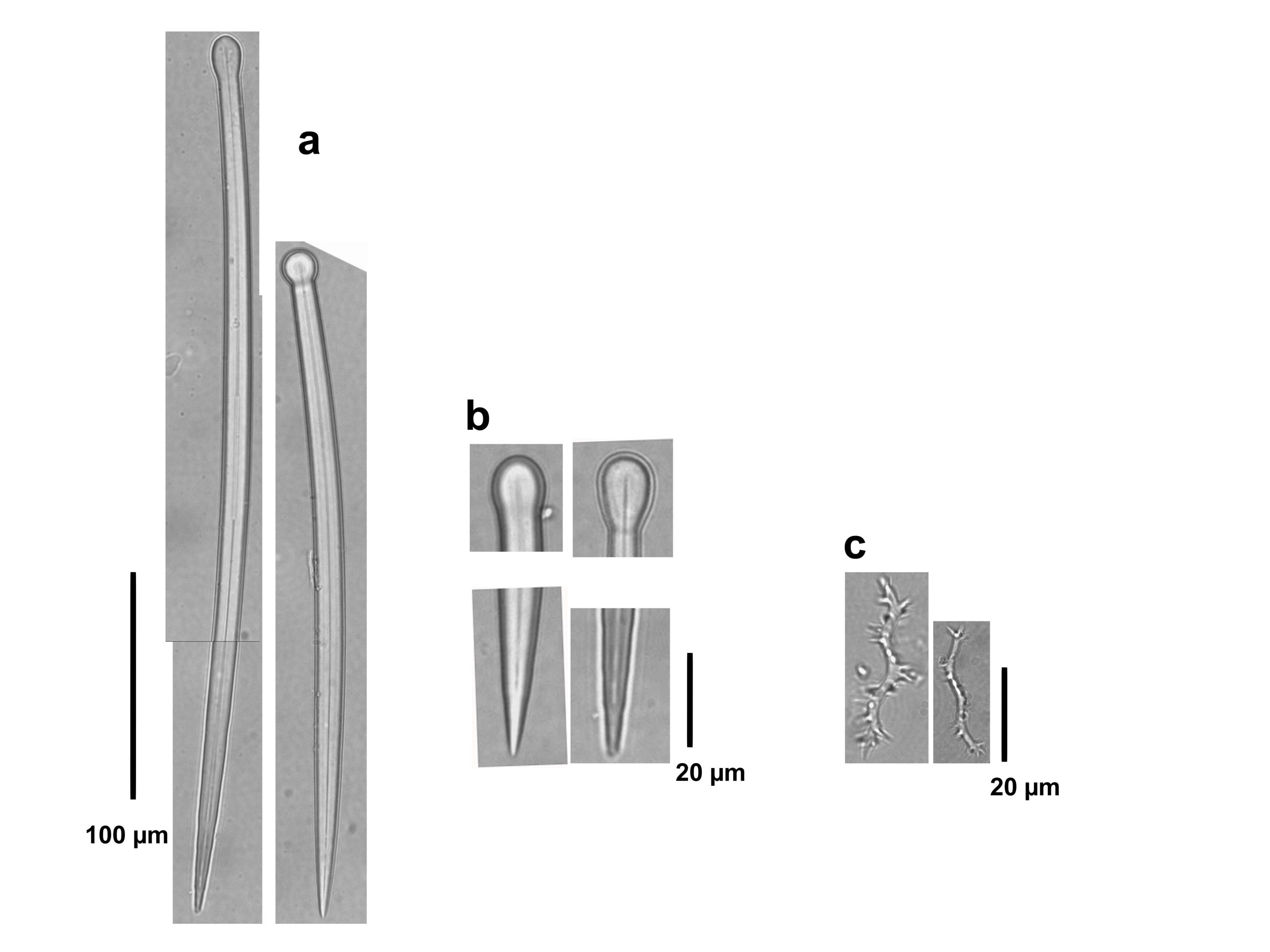
Description: Excavating, papillated sponge of low, thickly walled oscular papillae, separated from also low-lying inhalant ostial papillae, both up to 2 mm in diameter. Color of papillae yellow to orange yellow. Consistency soft. Megasclere spicules are robust tylostyles, 210-410 µm long by 4.5-18.8 µm wide, with rounded heads, often subterminal, 5-18 µm in width and 6.3-17.5 µm in length; ends acute. Microscleres are raphides, 97-375 µm long; no spirasters. The specimen from Santa Marta, Colombia, had the raphides, but the specimens from Bocas del Toro, Panama, and from Long Island, Bahamas lacked them. Tylostyles are more slender and thinner at Santa Marta (230-300 µm long and 4.5-7.3 µm wide), than at Bocas del Toro (210-410 µm long and 10-18.8 µm wide), following the generalized pattern of hypersilicification towards the Southern Caribbean.
Notes: The small size and the yellow color of the papillae, as well as the tylostyle shape and the presence of raphide microscleres fit the characteristics described for C. amplicavata from Bermuda (Rützler, 1974). However, we failed to find raphides in our material from the Bahamas and Colombia, although the other characteristics match the description. We decided to pool together these into C. amplicavata while further sampling yields more information about the variability of the species in the presence of microscleres. Other Caribbean excavating sponges with separated yellow papillae are C. janitrix sensu Pang, 1973, C. flavifodina Rützler, 1974, C. macgeachii Holmes, 2000, Siphonodityon brevitubulatum Pang, 1973, Alectona jamaicensis Pang, 1973, and Suberea flavolivescens (Hofman & Kielman, 1992), the latter three not yet pictured here. Apart from subtle differences in external shape, it is necessary to examine the spicules to distinguish them. Among those yellow Cliona mentioned above, species without spiraster microscleres are C. janitrix, and C. macgeachii. The former is distinguished from C. amplicavata by its tylostyles with abrupt stair-stepped ends and stylote and strongyloxeote modifcations, and the latter by its much smaller tylostyles (112-193 µm long by 3.6-10.4 µm wide).
Author Reference: Rützler, 1974
Link: World Porifera Database
Tissue and Spicule Images

Images
amplicavata
- Location: Panama-Bocas del Toro
- Photographer: Sven Zea

- Location: Panama-Bocas del Toro
- Photographer: Sven Zea
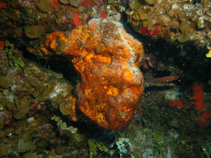
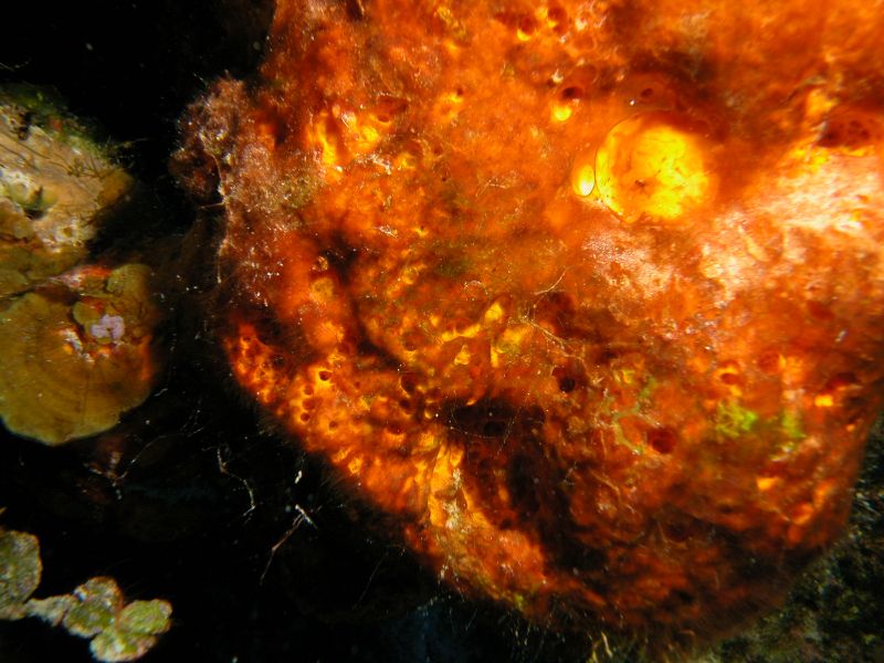
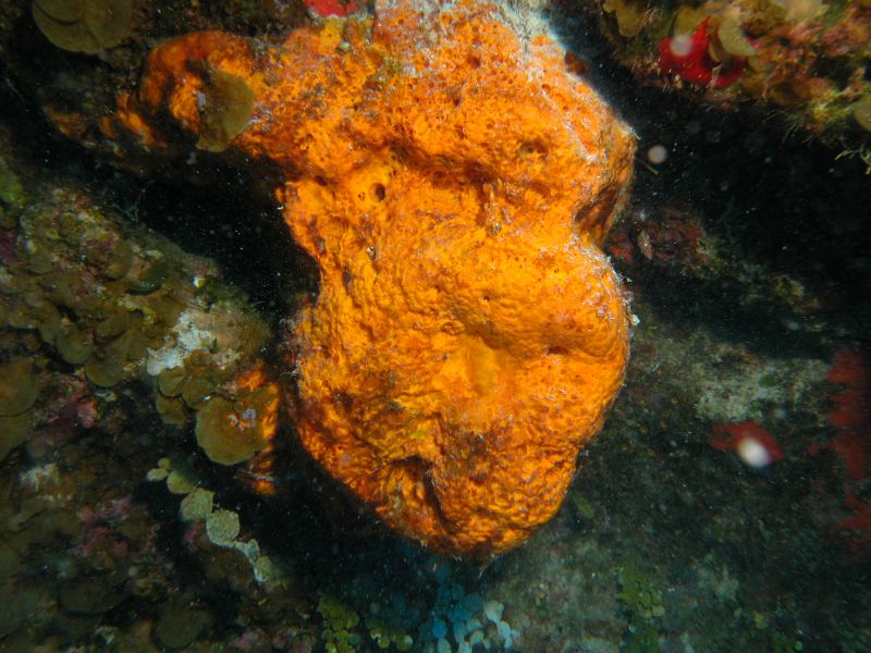
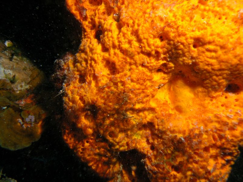
Observed Characteristics:
Color:
- orange
Morphology:
- papillated
Consistency:
- soft
Locations:
- Bahamas
Species Description and Notes
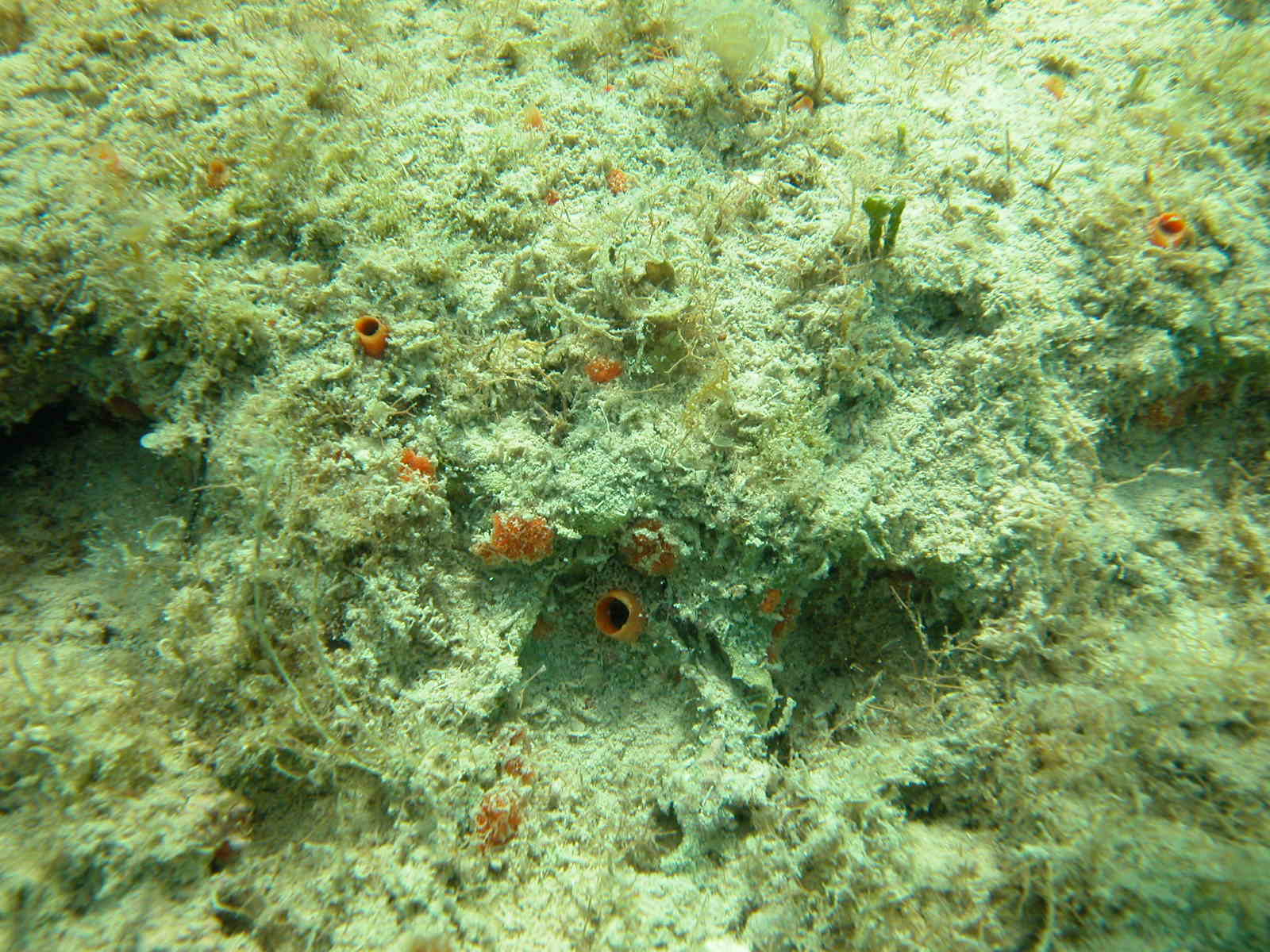
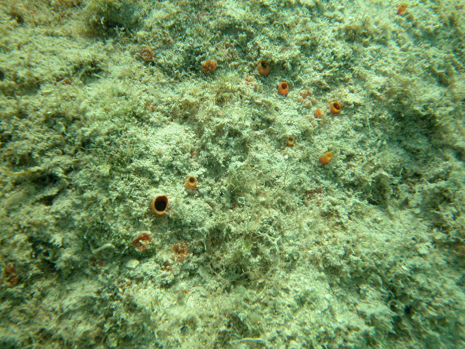
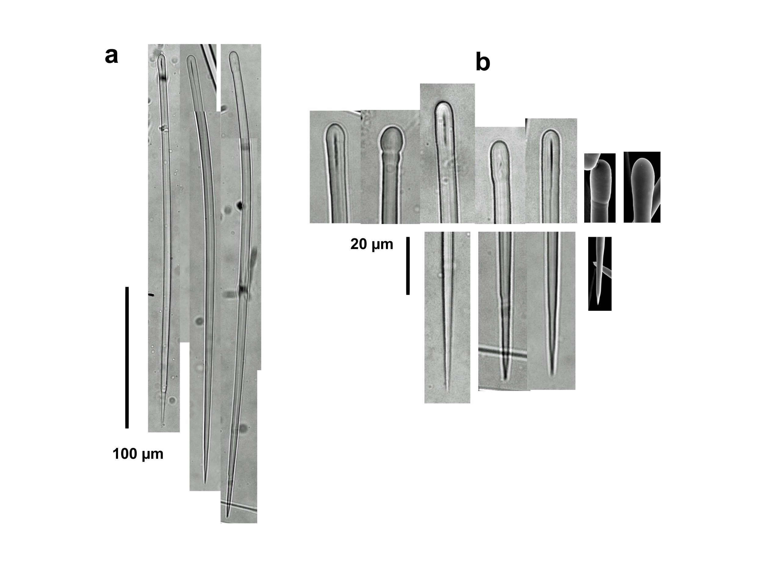
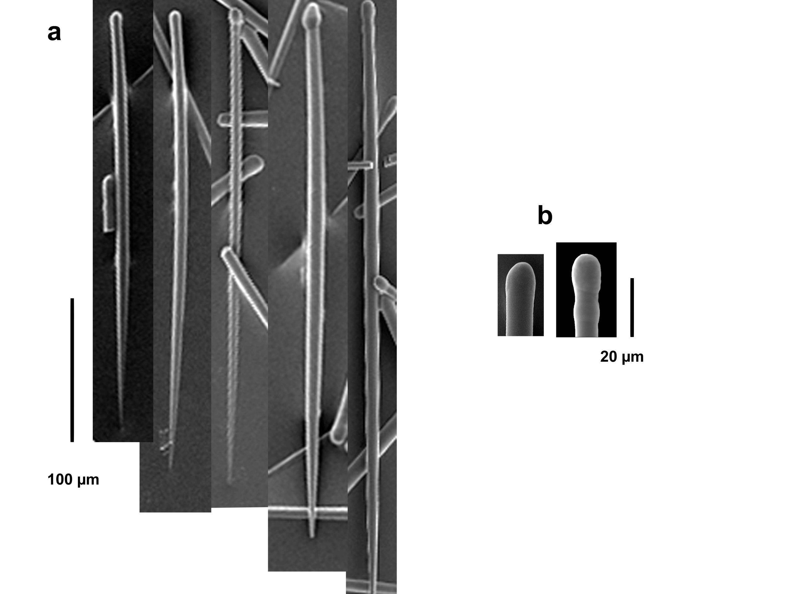
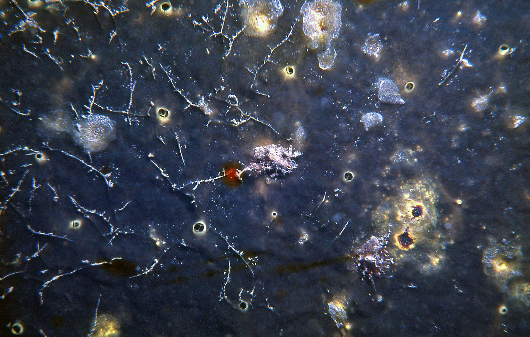
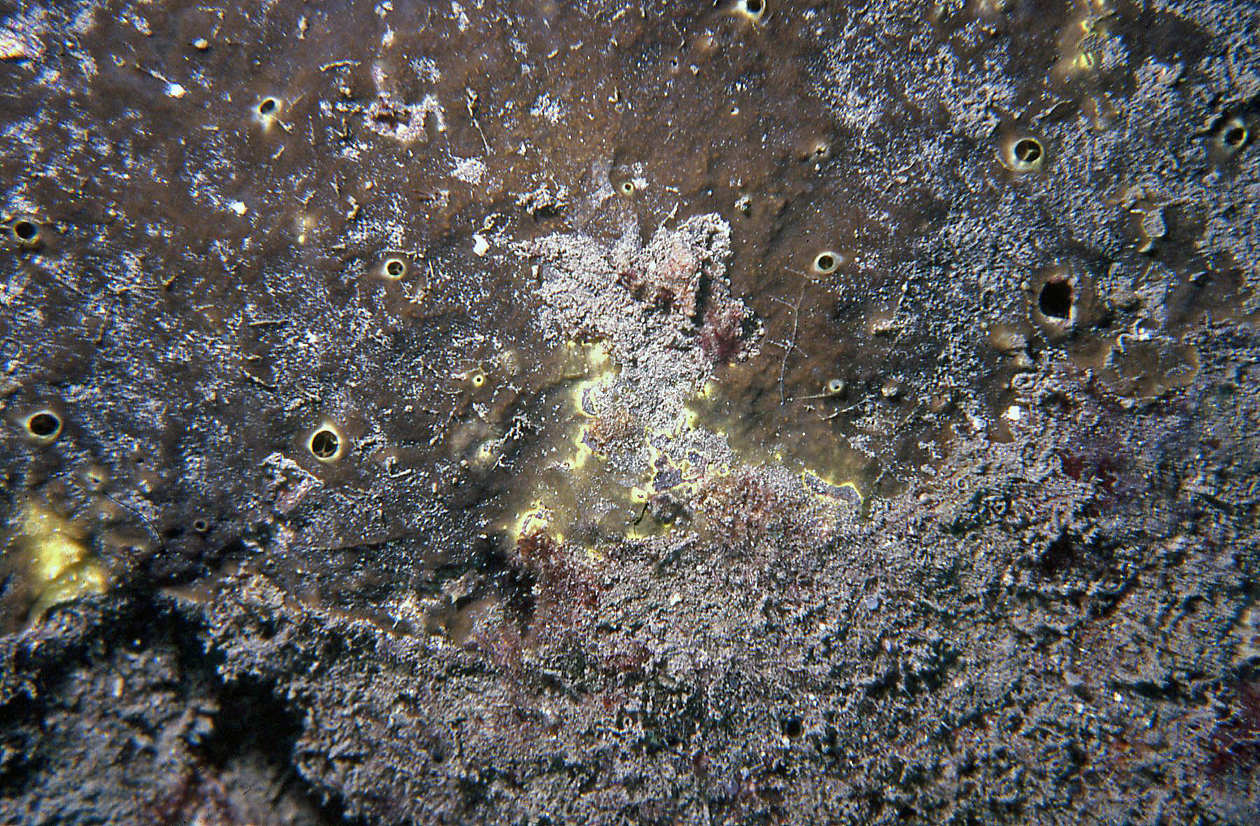
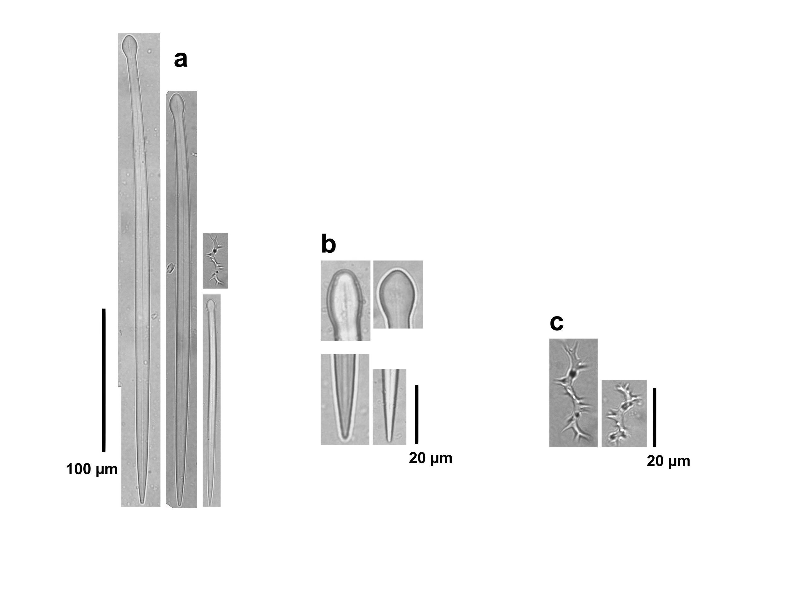
Description: Excavating, papillated sponge composed of separate oscular papillae surrounded by inhalant papillae, growing on exposed dead corals substratum or on bottom lagoon limestone, both heavily covered with algae and sediments. Papillae reach up to 5-10 mm in maximum diameter. Color orange. Consistency of epilythic portions soft. Skeleton of papillar tissues as palisades of spicules; deeper skeleton with many spicules in confusion. Megasclere spicules are slender to stout tylostyles with many stylote or strongyloxote modifications, 260-475 µm long by 3-14.6 µm wide; heads round and large, sometimes subterminal or deformed, 5-18 µm in diameter and 7.5-15.5 µm long; ends acute or mammiform, abruptly stair stepped; modifications include a few smooth styles or a rugose slight head, or a subterminal engrossing, or strongyloxoid spicules with both ends mammiform and asymmetric. No microscleres.
Notes: The identification of this species is tentative, C. janitrix sensu Pang, 1973 from Jamaica being the closest, owing to the irregular shape of the ends of the tylostyles and the presence of oxeote modifications with ends showing one or more abrupt reductions in diameter. However, the color of Jamaican specimens is dull yellow, contrasting with the orange from Bahamian specimens analyzed by us. Also, tylostyles are smaller and thinner in Jamaica, 134-221 µm long and 7.8-1 1.4 µm wide (Pang, 1973). The original C. janitrix is from the Mediterranean, so that even Pang (1973) record should be revised for the need of a new name. Specimens growing in sediment-laden lagoonal hard bottoms looked characteristically different to other Clionaids, but one specimen found dwelling in exposed coralline substratum was initially confused with papillated C. delitrix Pang, 1973, also pictured here, and only through the examination of spicules its identity was revealed.
Author Reference: Topsent, 1932
Link: World Porifera Database
Tissue and Spicule Images
Images
aff. janitrix
- Location: Bahamas-Sweetings Cay
- Photographer: Sven Zea
- Location: Bahamas-Sweetings Cay
- Photographer: Sven Zea
- Location: Bahamas-Sweetings Cay
- Photographer: Sven Zea
- Location: Bahamas-Sweetings Cay
- Photographer: Sven Zea
Observed Characteristics:
Color:
- brown
Morphology:
- encrusting
Consistency:
- tough
Locations:
- Colombia
Species Description and Notes
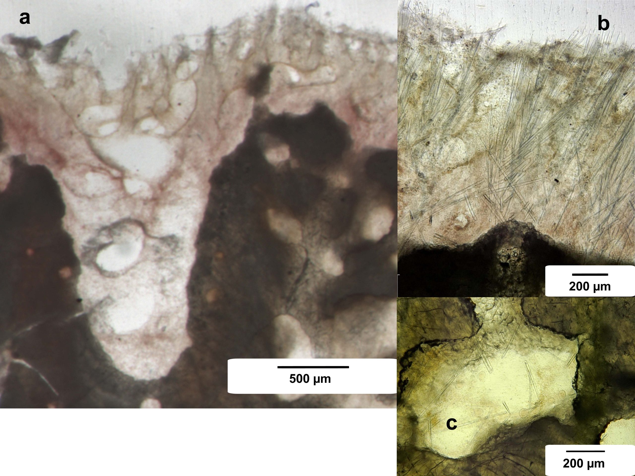
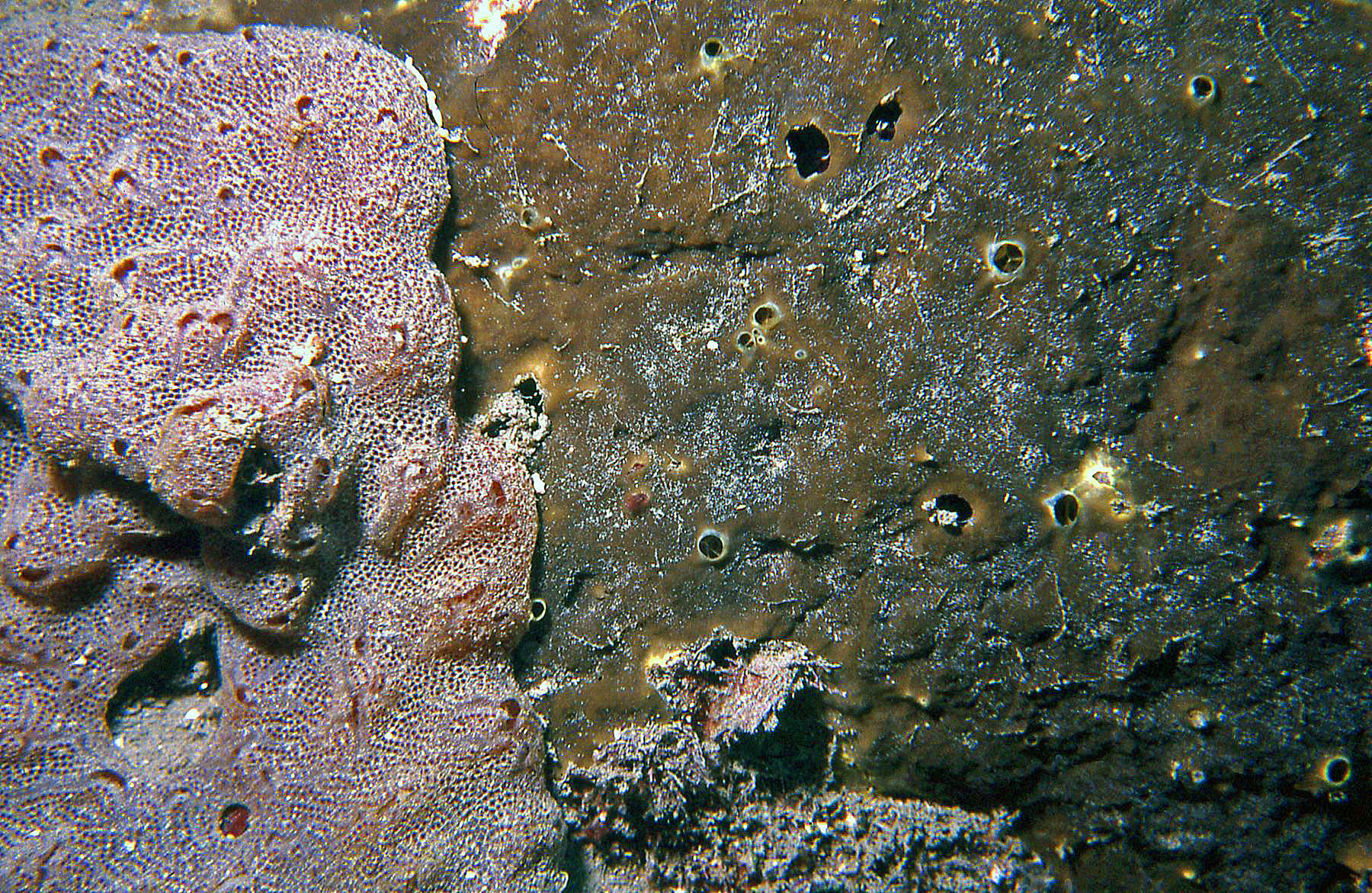
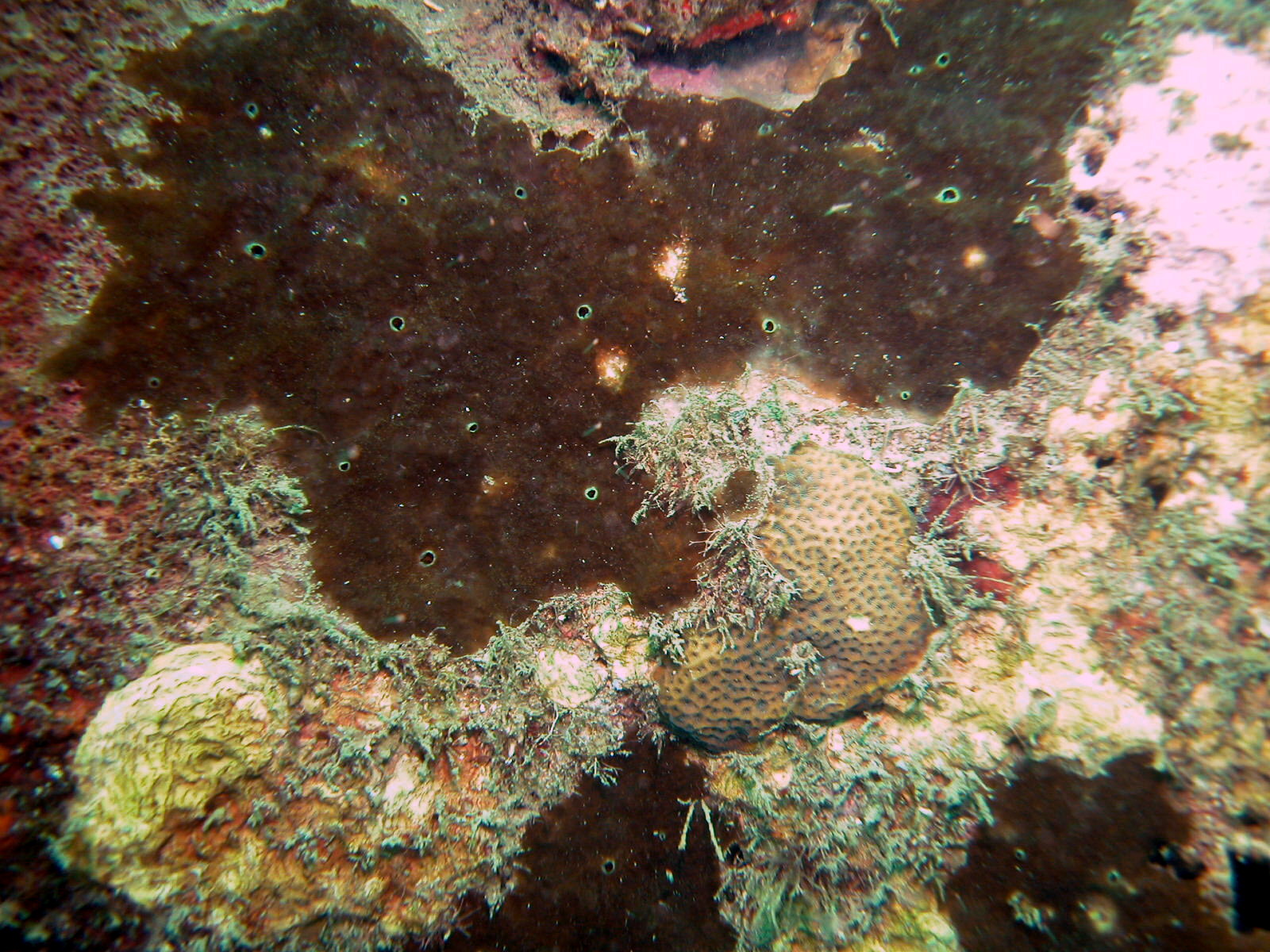
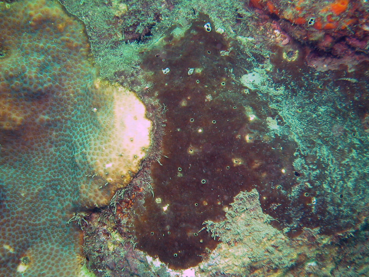
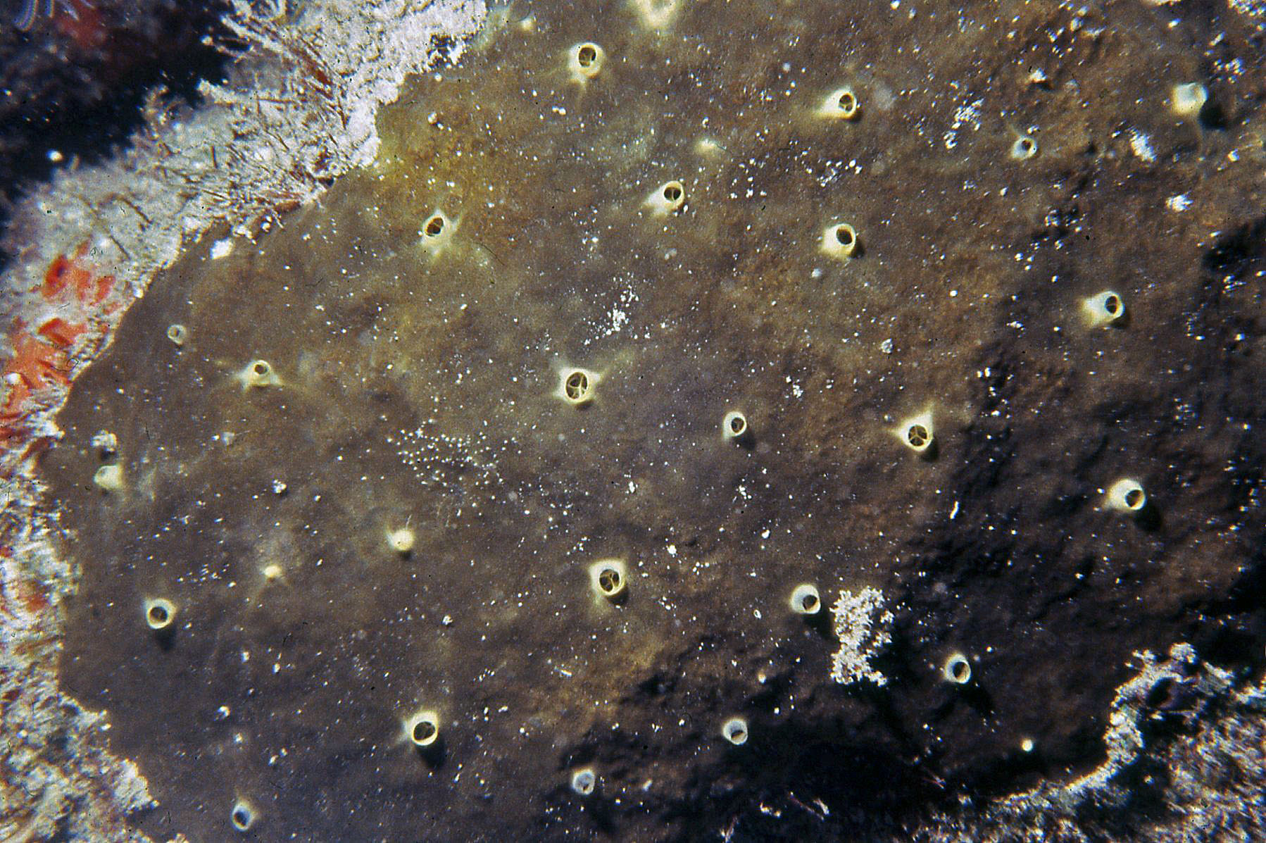
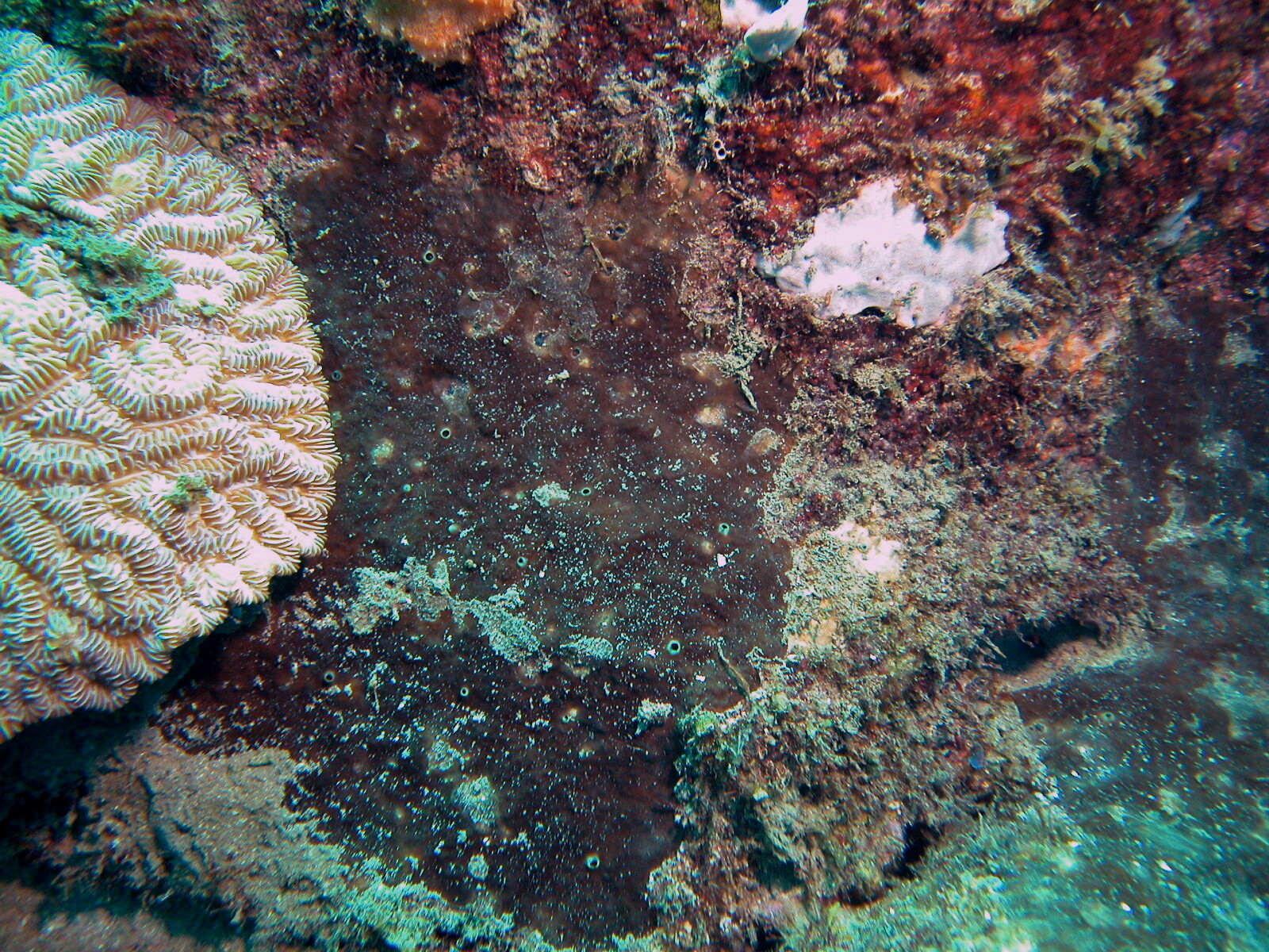
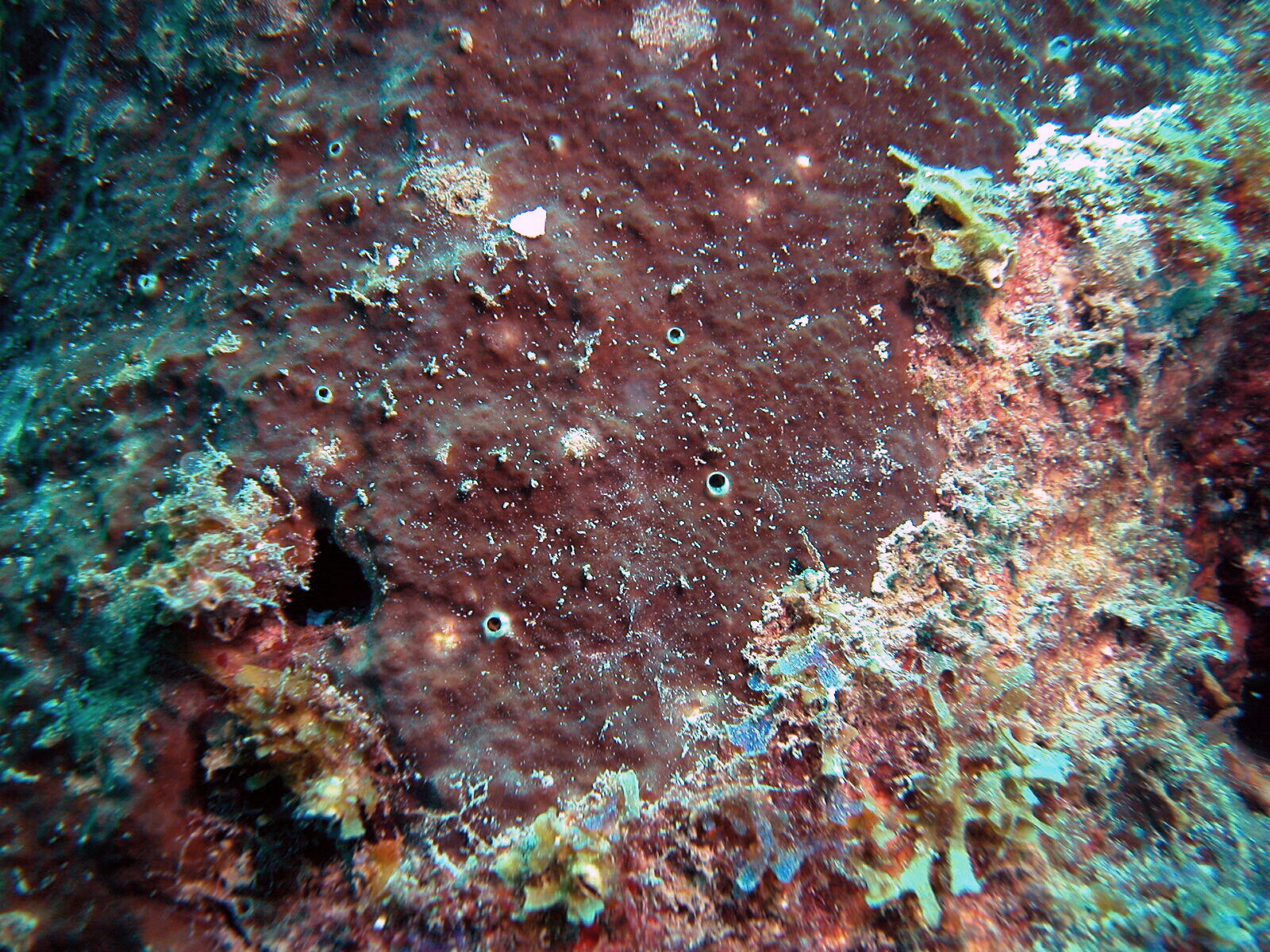
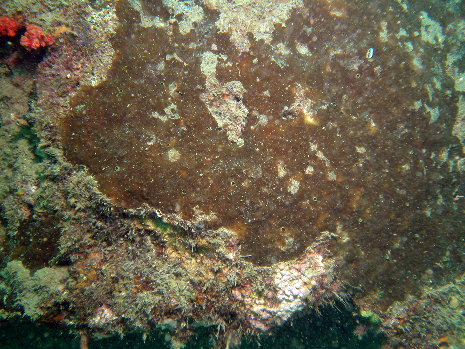
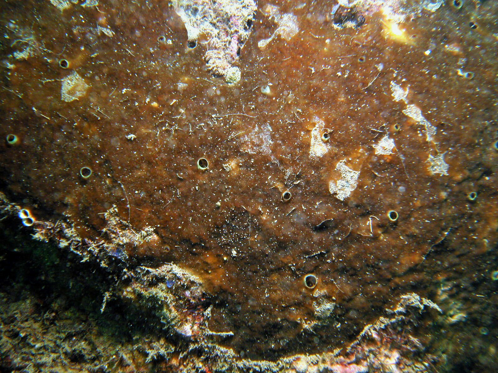
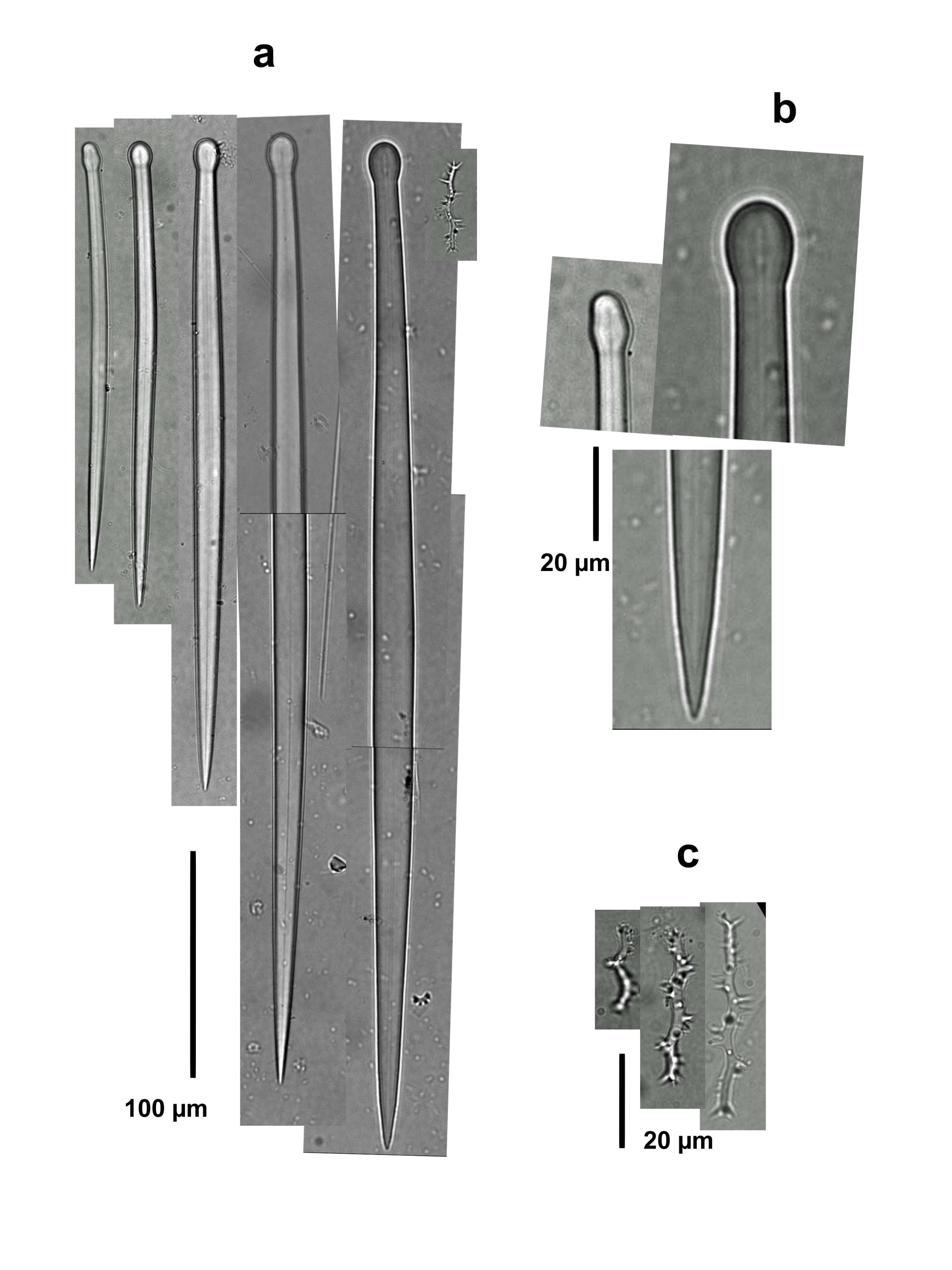
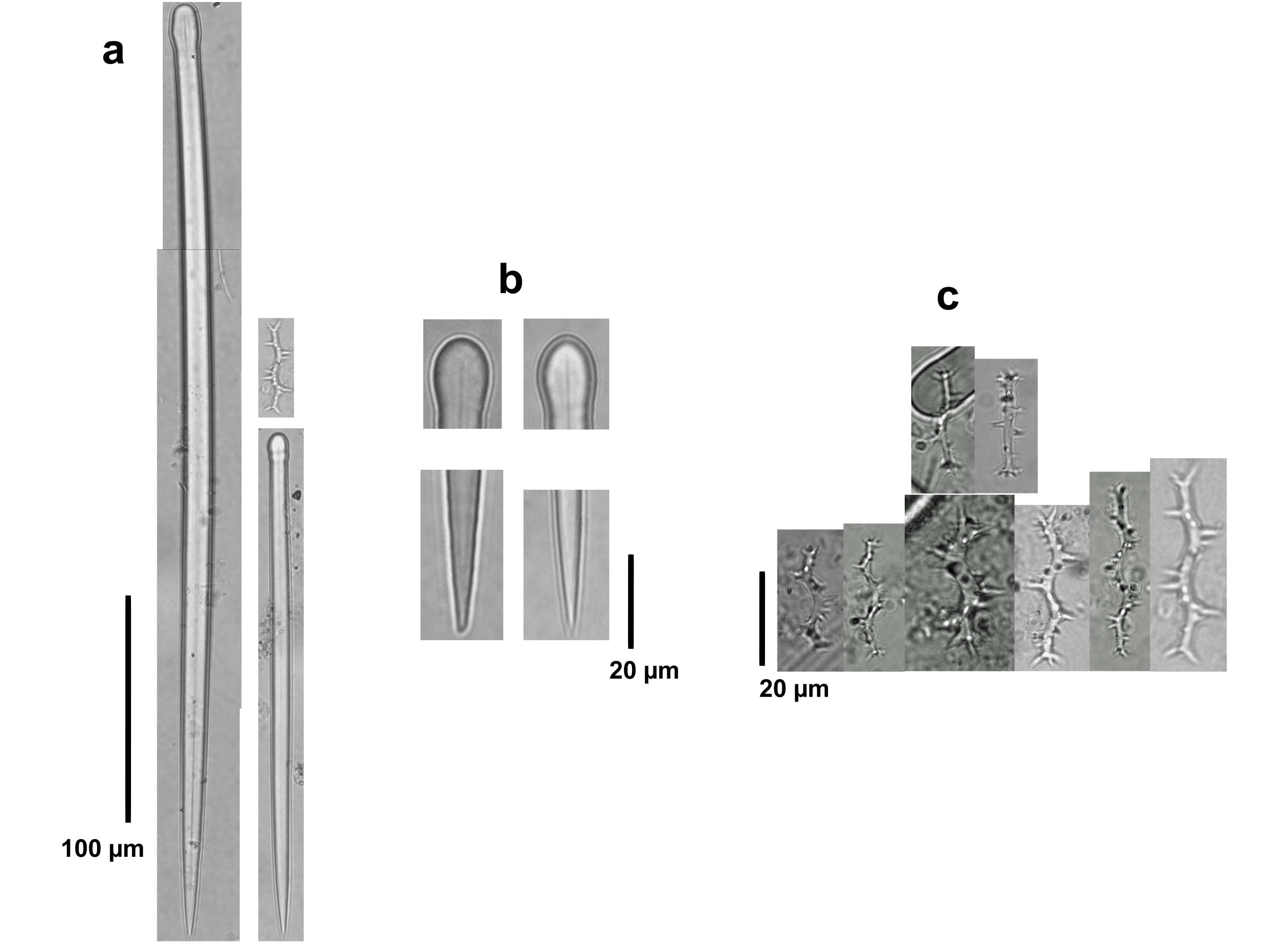
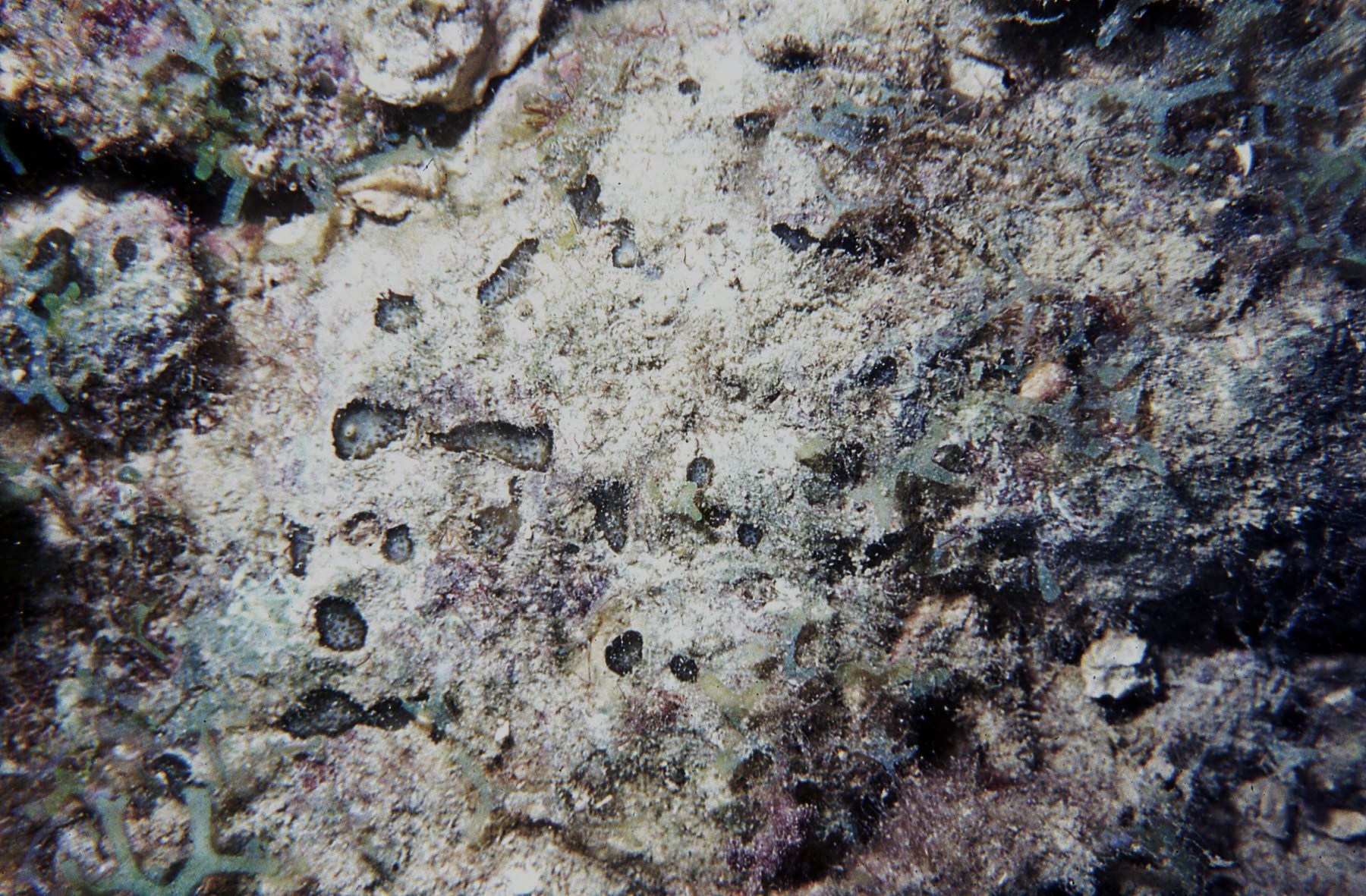
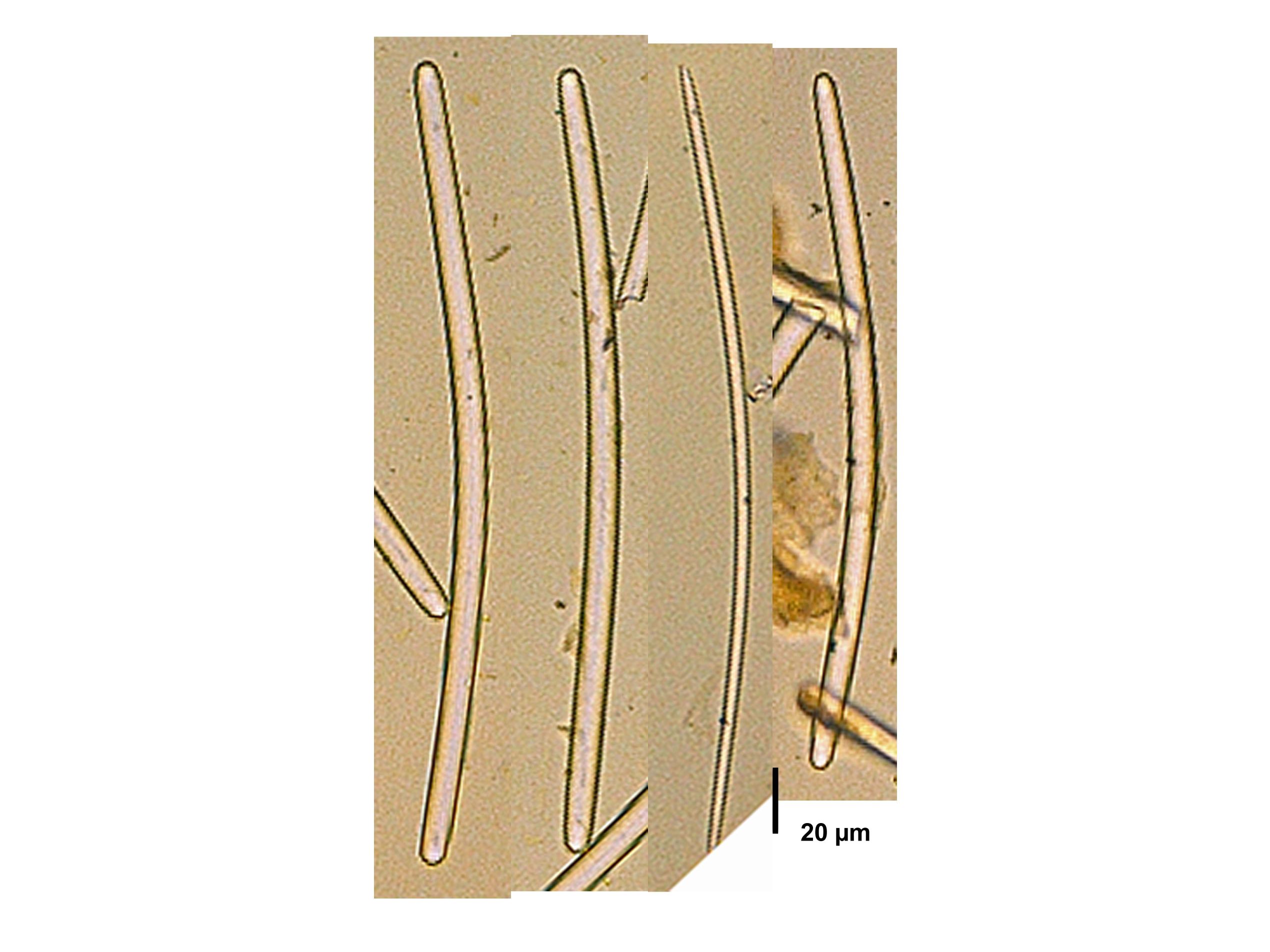
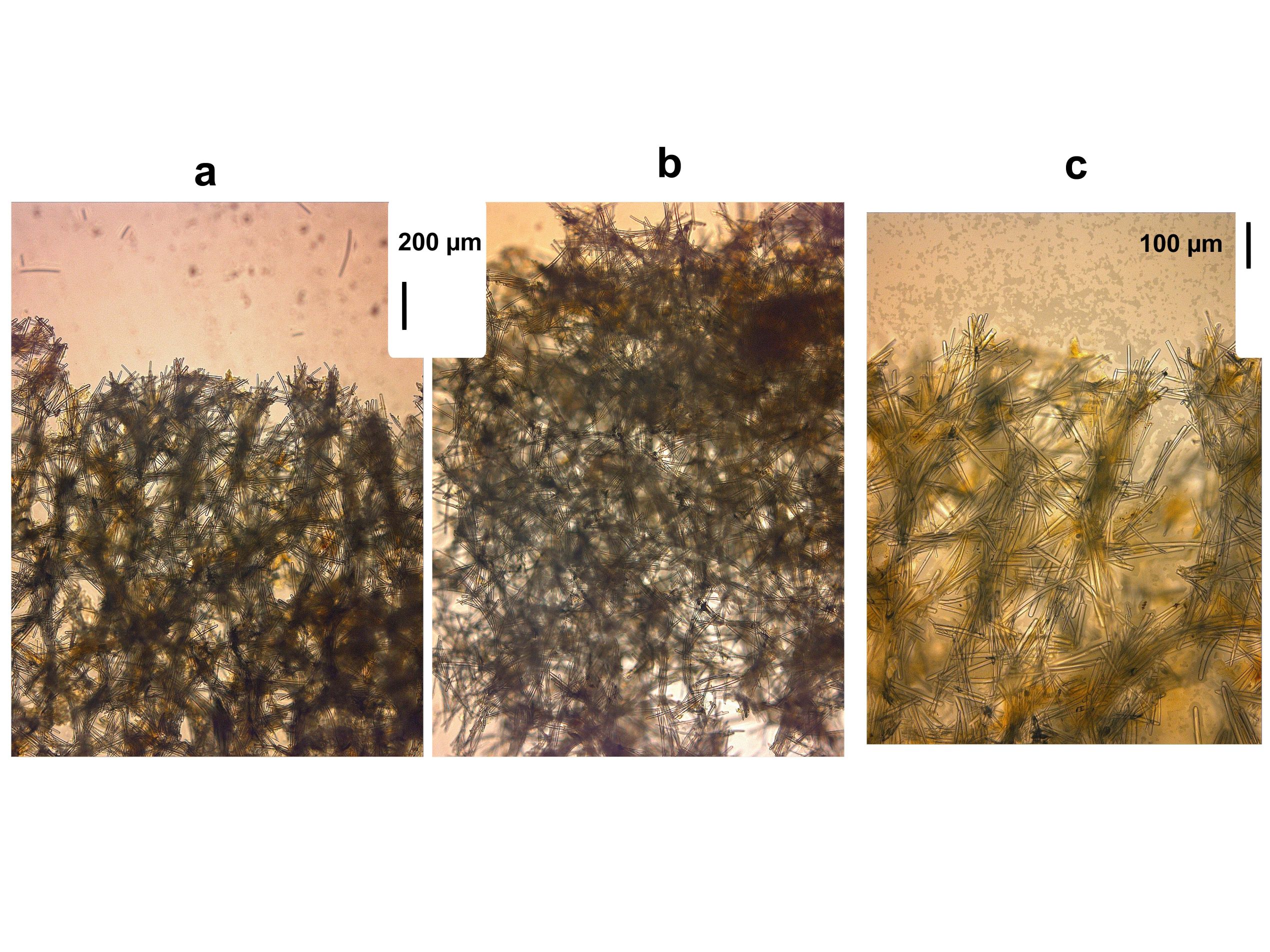
Description: (After Zea & López-Victoria, 2016) Excavating sponge, fully encrusting. It forms a round to irregular epilithic crust, 1-2 mm thick, and from a few cm to about 50-80 cm in maximum diameter. Oscules conspicuous, scattered, 1-8 mm in diameter, with a 1-2 mm tall membranous collar. Surface color amber brown to gray brown, sometimes greenish; oscular collars bright sulfur yellow. Consistency of the ectosome leathery, crumbly for the choanosomal tissue. Surface as even, or as rugose as the underlying substratum; smooth, rather velvety, shiny when not covered by sediment-ladden mucus. Sponge penetrates and excavates about 1-3 cm underneath the surface. Excavating tissue ochre yellow, fills calicular spaces and excavated chambers. Ectosomal skeleton as erect, tree-like plumose tracts up to 8–10 spicules thick, separated at the base 200–300 μm, which ascend and divide 1–2 times up to the surface. Choanosomal skeleton of ill-defined tracts of 2–3 spicules, more often located towards the walls of chambers. Chamber walls creased with concave erosion scars, up to 60 μm wide. Megascleres spicules tylostyles, slender, slightly curved, 280-480 μm long and 5.8-17.5 μm wide; some tylostyles lack a head, but for most the heads are narrow, elongated, more bulged on one side, often misshaped, some subterminal; tips long, acute, most often telescoped. Heads, when present, 6.9-10.3 μm wide and 8.1-15.0 μm long. Microsclere spirasters absent.
Notes: Other Caribbean dark brown fully encrusting excavating sponges of the genus Cliona reaching large sizes are C. caribbaea Carter, 1882, C. tenuis Zea & Weil, 2003 and C. varians (Duchassaing & Michelotti, 1864), all pictured here. C. caribbaea and C. tenuis always possess spiraster microscleres (lacking in C. acephala) and the heads of the tylostyles are always clearly delineated and rarely deformed, usually drop-shaped to rounded, in contrast to the narrow to deformed or absent heads in C. acephala. Tylostyle dimensions of C. acephala (280–480 x 5.8–17.5 μm) overlap with those of C. caribbaea (271–418 x 4.7–15.2 μm) but are larger than in C. tenuis (199–380 μm x 3.3–14.3 μm) (Zea & Weil 2003). While C. caribbaea can occur in papillate morphology, its encrusting form is like that of C. acephala and develops a similar thickness of the epilithic crust and has a similar oscular diameter, but the tylostyle shape is very different between the two species (Zea & Weil 2003). Although reef-dwelling specimens of C. varians are usually encrusting (also called forma incrustans Wiedenmayer, 1977) and tan to yellow brown, the few that are dark brown can potentially be confused in the field with C. acephala. C. varians is distinguished by a greater thickness of the epilithic crust (up to 3–6 mm), much larger oscules (up to 1 cm in diameter) and its characteristic C-shaped spirasters (also called anthosigmas). Phylogenetic analysis of nuclear ribosomal internal transcribed spacer 2 (ITS2) DNA sequences carried out by Escobar et al. (2012) showed C. acephala as genetically distinct from the other Caribbean congeners, and more closely related to Indo-Pacific C. orientalis Thiele, 1900.
Author Reference: Zea & López-Victoria, 2016
Link: World Porifera Database
Tissue and Spicule Images
Images
acephala
- Location: Colombia-Santa Marta
- Photographer: Sven Zea
- Location: Colombia-Santa Marta
- Photographer: Sven Zea
- Location: Colombia-Santa Marta
- Photographer: Sven Zea
- Location: Colombia-Santa Marta
- Photographer: Sven Zea
- Location: Colombia-Santa Marta
- Photographer: Sven Zea
- Location: Colombia-Santa Marta
- Photographer: Sven Zea
- Location: Colombia-Santa Marta
- Photographer: Sven Zea
- Location: Colombia-Santa Marta
- Photographer: Sven Zea
- Location: Colombia-Santa Marta
- Photographer: Sven Zea
- Location: Colombia-Santa Marta
- Photographer: Sven Zea
- Location: Colombia-Santa Marta
- Photographer: Sven Zea
- Location: Colombia-Santa Marta
- Photographer: Sven Zea
- Location: Colombia-Santa Marta
- Photographer: Sven Zea
- Location: Colombia-Santa Marta
- Photographer: Sven Zea
Observed Characteristics:
Color:
- cinnamon-tan
- red
- brown
- white with brown edges
Morphology:
- papillated
Consistency:
- tough
Locations:
- Colombia
Species Description and Notes
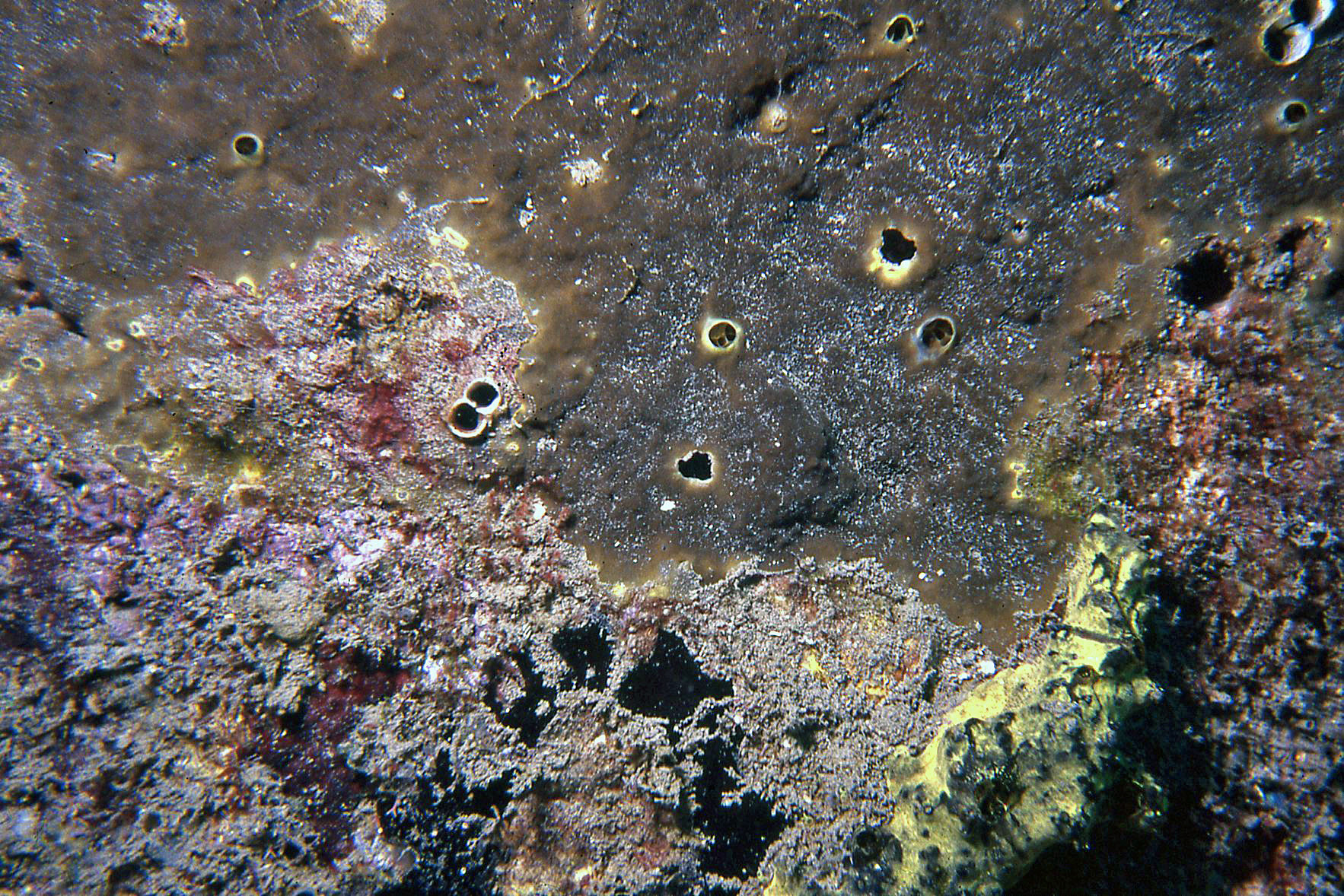
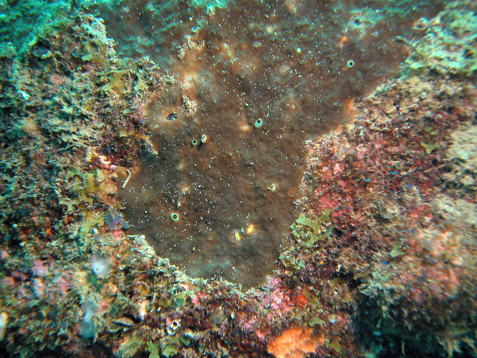
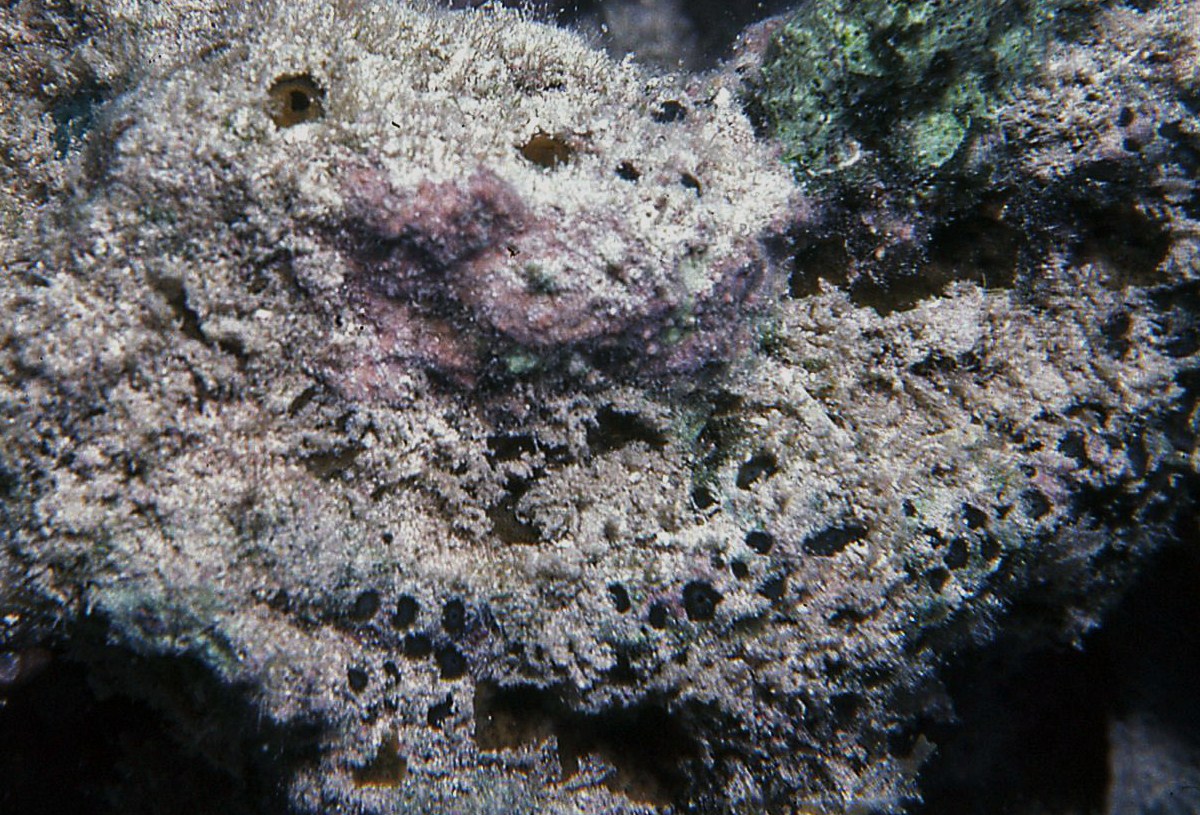
Description: Excavating sponge, showing low-lying papillae, scattered on the coralline substratum, light brown to tan to whitish (with brownish edges) in color. Papillae also reported red. Oscular and inhalant papillae are usually separated. Oscular papillae up to about 1 cm in diameter, visible as a thick tissue collar surrounding the osculum; inhalant papillae irregular and ragged in outline, up to several cm in maximum diameter, typically depressed towards the center, pierced by pores. When the substratum is broken, the galleries are wide (up to 1-2 cm in diameter), lined with yellowish tissue. Megasclere spicules are tylostyles, slightly curved, with drop-shaped heads and smooth, pointed ends, 180-430 µm long by 6.2-12.5 µm wide; the heads are 8.8-17 µm wide by 9.5-18 µm long. Microsclere spicules are spirasters, 15-50 µm long, slender, with a shaft up to about 2-3 µm thick, with up to 3-4 relatively narrow (6 µm wide) turns; spines are characteristically long and usually not divided.
Notes: This species lives in shallow reef and rocky locations. What was described as Cliona caribbaea by Pang (1973), turned out to be this species (see Zea & Weil, 2003). Also, some of the specimens named C. caribbaea by Hofman & Kielman (1992) from Santa Marta, Colombia are C. flavifodina. This species is distinguished from other Caribbean species by its wide and irregular inhalant papillae, and the longish spines of the spirasters.
Author Reference: Rutzler, 1974
Link: World Porifera Database
Tissue and Spicule Images
Images
flavifodina
- Location: Colombia-Islas de San Bernardo
- Photographer: Sven Zea
- Location: Colombia-Islas del Rosario
- Photographer: Sven Zea
Observed Characteristics:
Color:
- brown
Morphology:
- branching
Consistency:
- tough
Locations:
- Dominican Republic
Species Description and Notes
Description: Transcribed from Pulitzer-Finali (1986): “A small fragment is available. The sponge was subcylindrical, 2.5-3 cm in diameter, Brown, firmly resilient, densely infested by a zoanthid. In the dry state it has the consistency of cork. Oscules are flush, sparse, 2 mm wide. At the surface the skeleton is a not separable reticulation of spicular tracts which form not quite distinct meshes 350 µm wide. Around the perforations made by the zoanthid there is a collar of spicules accumulated in confusion. The choanosomal reticulation of irregular tracts forming meshes of 500-700 µm is obscured by a large number of spicules in disorder. Spicules: strongyles slightly curved, measuring 140-260 x 5-14 µm. No distinct categories are recognizable.”
Notes: This record is here included thanks to having photos of the spicules and skeleton of the type. They were used to make comparisons with three species-morphs included here under the genus Amphimedon?, that share the same type of spicules, but a much looser skeletal arrangement. This species has not been reported after the original description and its status is in need to be defined.
Author Reference: (Pulitzer-Finali, 1986)
Link: World Porifera Database
Tissue and Spicule Images
Images
dominicana
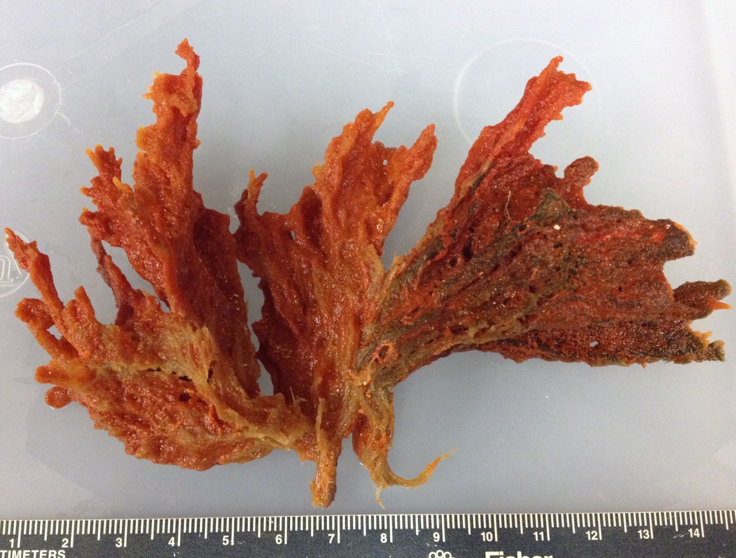
Observed Characteristics:
Color:
- orange
Morphology:
- bushy
- fan
Consistency:
- tough
Locations:
- United States
Species Description and Notes
Notes: Orange-red bushes with fan-like folds. Could be confused with other bushy species with similar color such as Monanchora arbuscula (Duchassaing & Michelotti, 1864), also pictured here. Florida and Gulf of Mexico records of Clathria prolifera (Ellis & Solander, 1786) may be attributed to this species, which lives further north in the western Atlantic. For a redescription see Gómez (2014).
Author Reference: Topsent, 1889
Link: World Porifera Database
Tissue and Spicule Images
Images
(Clathria) foliacea
- Location: United States, Florida Keys
- Photographer: Joseph Pawlik

- Location: United States, Florida Keys
- Photographer: Sven Zea
- Location: United States, Florida Keys
- Photographer: Sven Zea
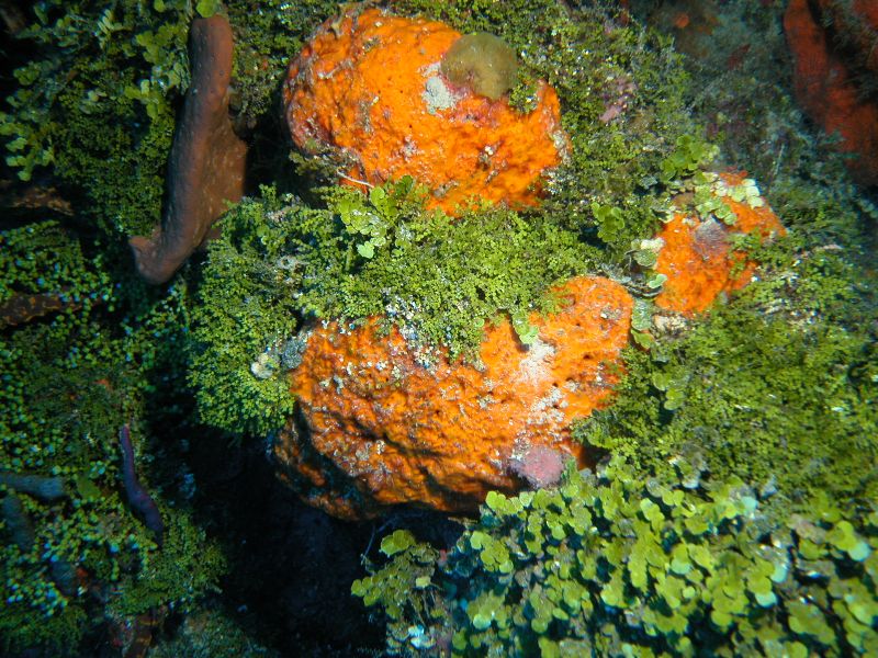
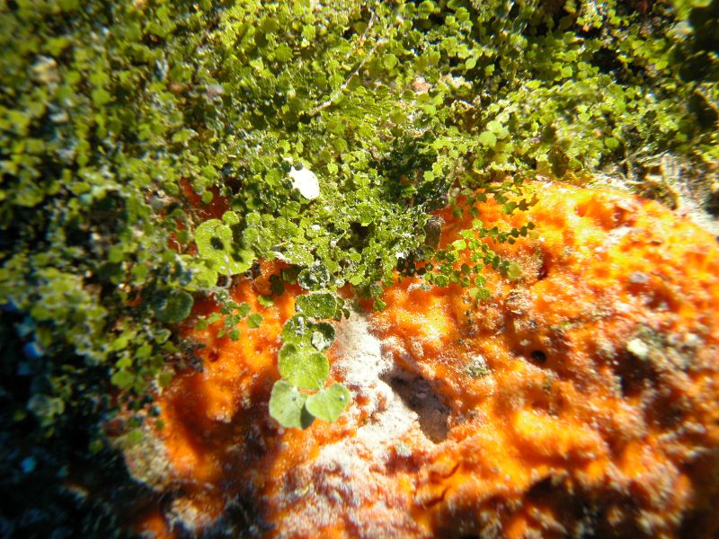
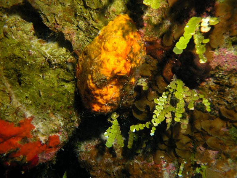
Observed Characteristics:
Color:
- orange
- orange-yellow
Morphology:
- massive
- spherical
Consistency:
- crumbly
Locations:
- Bahamas
Species Description and Notes
Notes: Orange-yellow irregular masses with rugose surface, often growing among algae in vertical cliffs; crumbly. It may be confused with massive or cavity-filling specimens of Agelas citrina Alcolado, 1987, which are tougher.
Author Reference: Lehnert & van Soest, 1996
Link: World Porifera Database
Tissue and Spicule Images
Images
(Clathria) faviformis
- Location: Little San Salvador, Bahamas
- Photographer: Sven Zea
- Location: Little San Salvador, Bahamas
- Photographer: Sven Zea
- Location: San Salvador, Bahamas
- Photographer: Sven Zea
- Location: San Salvador, Bahamas
- Photographer: Sven Zea
- Location: San Salvador, Bahamas
- Photographer: Sven Zea
- Location: San Salvador, Bahamas
- Photographer: Sven Zea
- Location: San Salvador, Bahamas
- Photographer: Sven Zea

- Location: San Salvador, Bahamas
- Photographer: Sven Zea
Observed Characteristics:
Color:
- red
- orange
Morphology:
- encrusting
Consistency:
- soft
Locations:
- Bahamas
Species Description and Notes
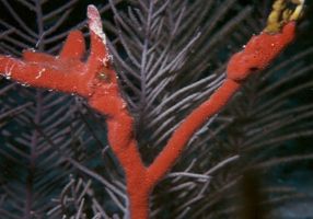
Notes: Thinly encrusting bright red orange, with slighlty evident exhalant canals in a star pattern. The spiculation consists of stout, curved tylostyles 350-480 x 10-15 µm, straight subtylostyles 275-450 µm, toxa bulging towards the center (hence the name bulbotoxa) 45-180 µm, and twisted isochelae 12-15 µm. It is difficult to distinguish it in the field from other bright orange encrusting sponges such as Clathria spinosa Wilson, 1902 or Placospherastra micraster (Lehnert & Heimler, 2001), both pictured here.
Author Reference: van Soest, 1984
Link: World Porifera Database
Tissue and Spicule Images
Images
(Microciona) bulbotoxa
- Location: Bahamas, Hogsty Reef
- Photographer: Sven Zea
Observed Characteristics:
Color:
- red
- orange
Morphology:
- encrusting
Consistency:
- soft
Locations:
- Bahamas
Species Description and Notes
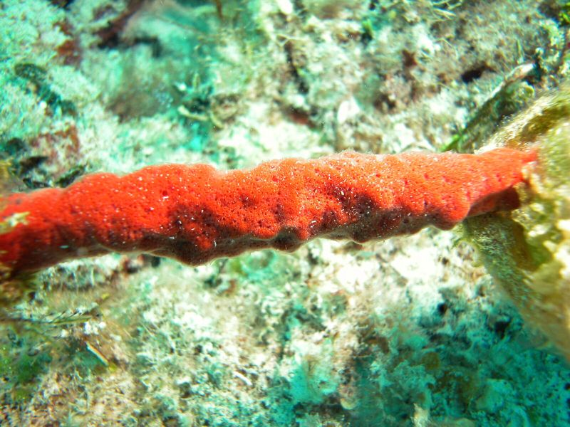
Notes: Red, thick encrustation on gorgonian branches. Surface looks pierced by numerous, contiguous perforations. Transparent skin outlines oscules and exhalant canals.
Author Reference: (de Laubenfels, 1934)
Link: World Porifera Database
Tissue and Spicule Images
Images
(Microciona) calla
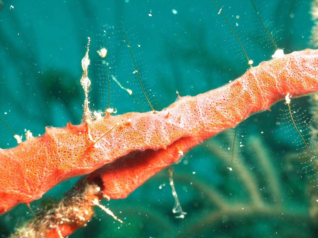
- Location: Bahamas, Sweetings Cay
- Photographer: Sven Zea
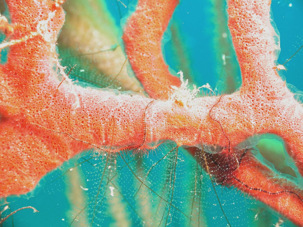
- Location: Bahamas, Sweetings Cay
- Photographer: Sven Zea
- Location: Bahamas, Stirrups Cays, N Berry Islands
- Photographer: Sven Zea
- Location: Bahamas, Stirrups Cays, N Berry Islands
- Photographer: Sven Zea
- Location: Bahamas, Stirrups Cays, N Berry Islands
- Photographer: Sven Zea