
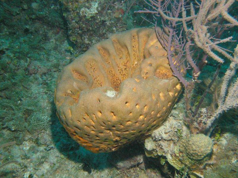
- Location: Sweetings Cay, Bahamas
- Habitat: deep reef
- Photographer: Sven Zea
- Picture Taken On: 2007-06-17 21:52:39
- Picture Notes: no image notes
- Color: cinnamon-tan
- Morphology: vase
- Consistency: hard
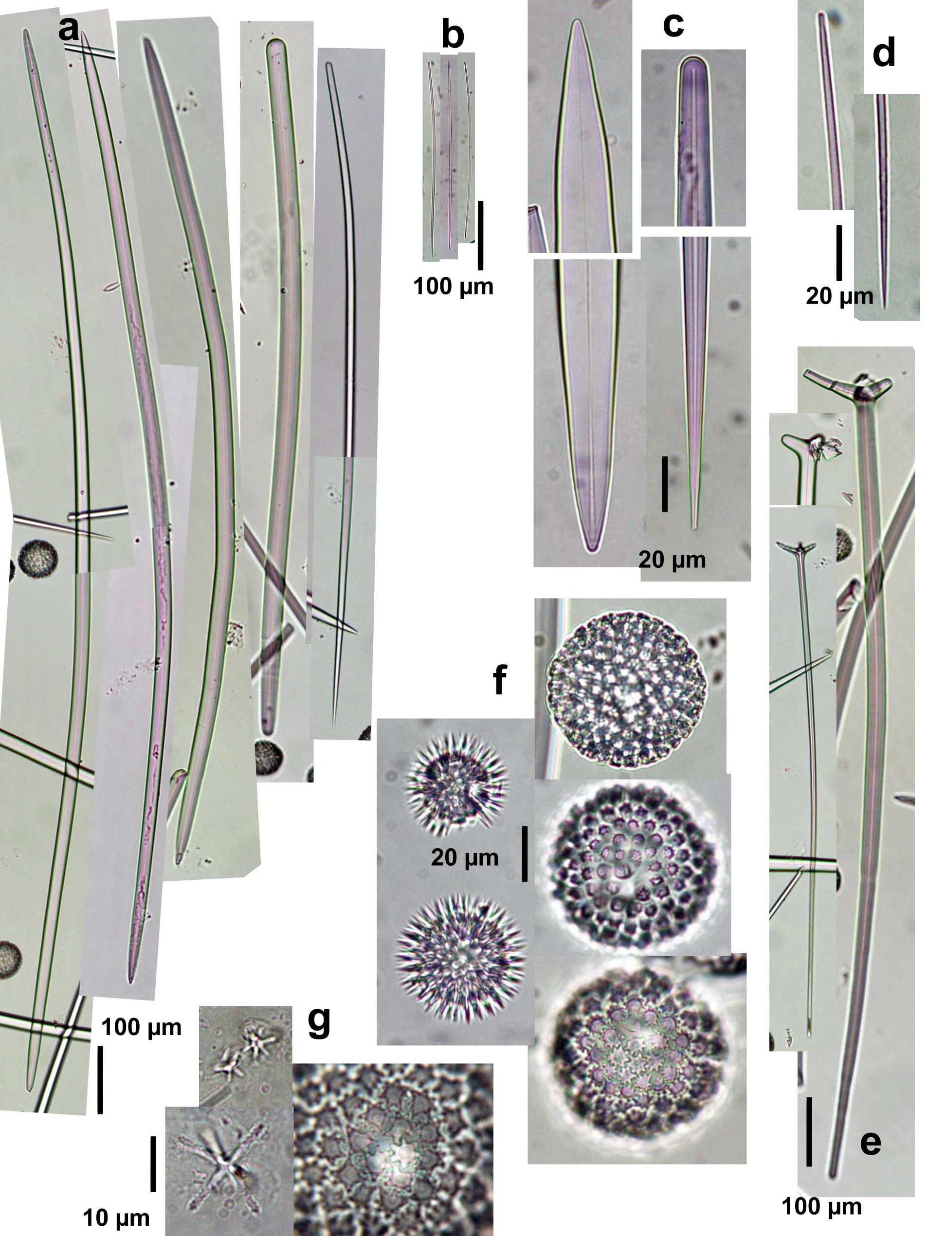
Spicules Image: a) Oxeas-styloids-styles I; b) oxeas-anysostrongyloxeas II, c) enlarged tips of oxeas-styloids-styles I; d) enlargement of oxeas-anysostrongyloxeas II; e) plagiotriaenes; f) microscleres (sterrasters, developed and developing); g) enlargement of microscleres (rossettes of sterrasters on the right; spined asters in 2 types, oxyasters and and strongyl- or oxyasters). Sample from the Bahamas.
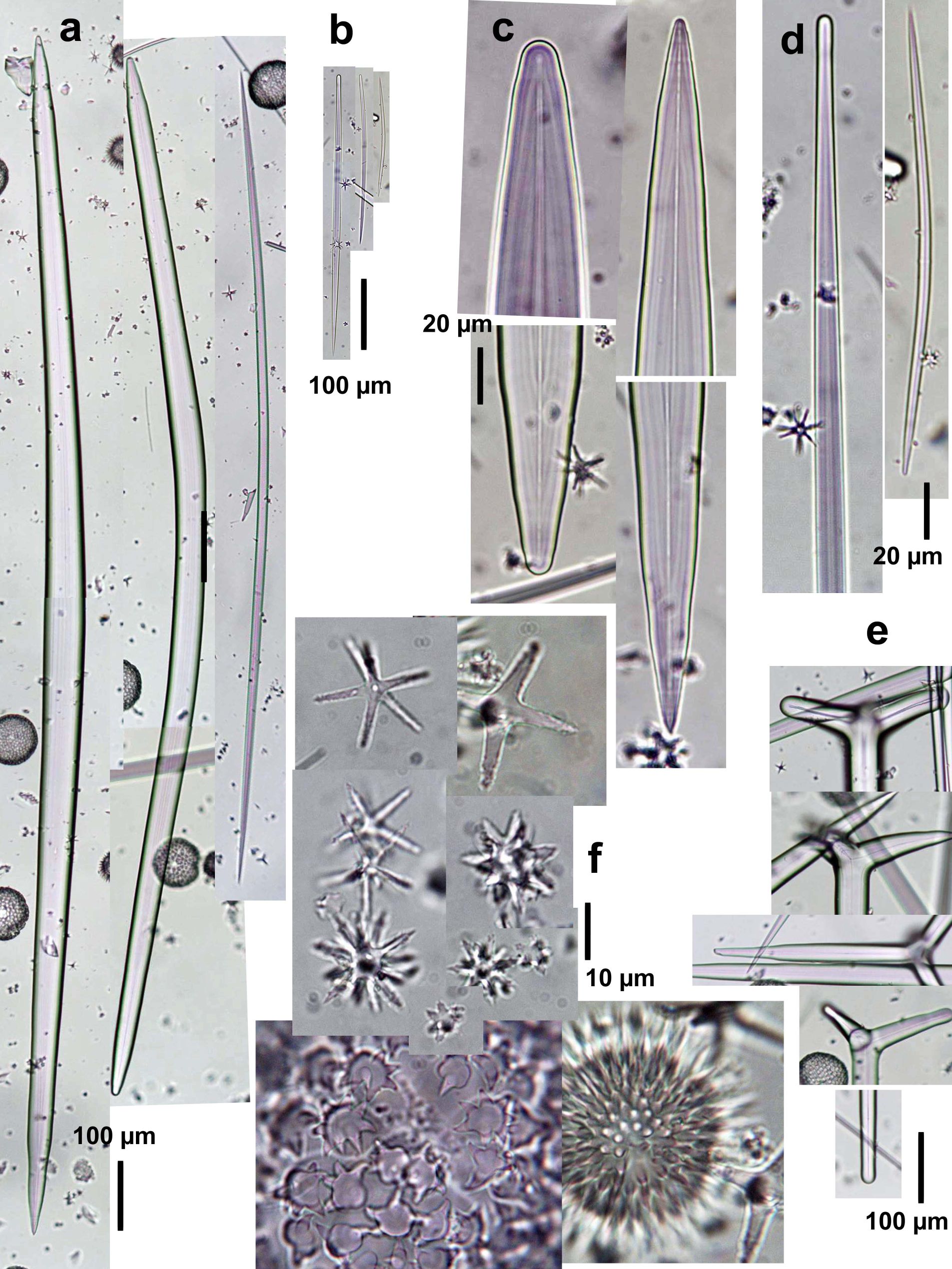
Spicules Image: a) Oxeas I with sterrasters; b) anysostrongyloxeas-oxeas II, c) enlarged tips of oxeas I; d) enlargement of anysostrongyloxeas-oxeas II; e) plagiotriaenes; f) enlargement of microscleres (rossettes of sterrasters on the bottom; spined asters in 2-3 types, oxyasters and strongyl- or oxyasters). Sample from Islas del Rosario, Colombia. Notice the larger and thicker megascleres and stouter microscleres in comparison to those pictured in a sample from the Bahamas.
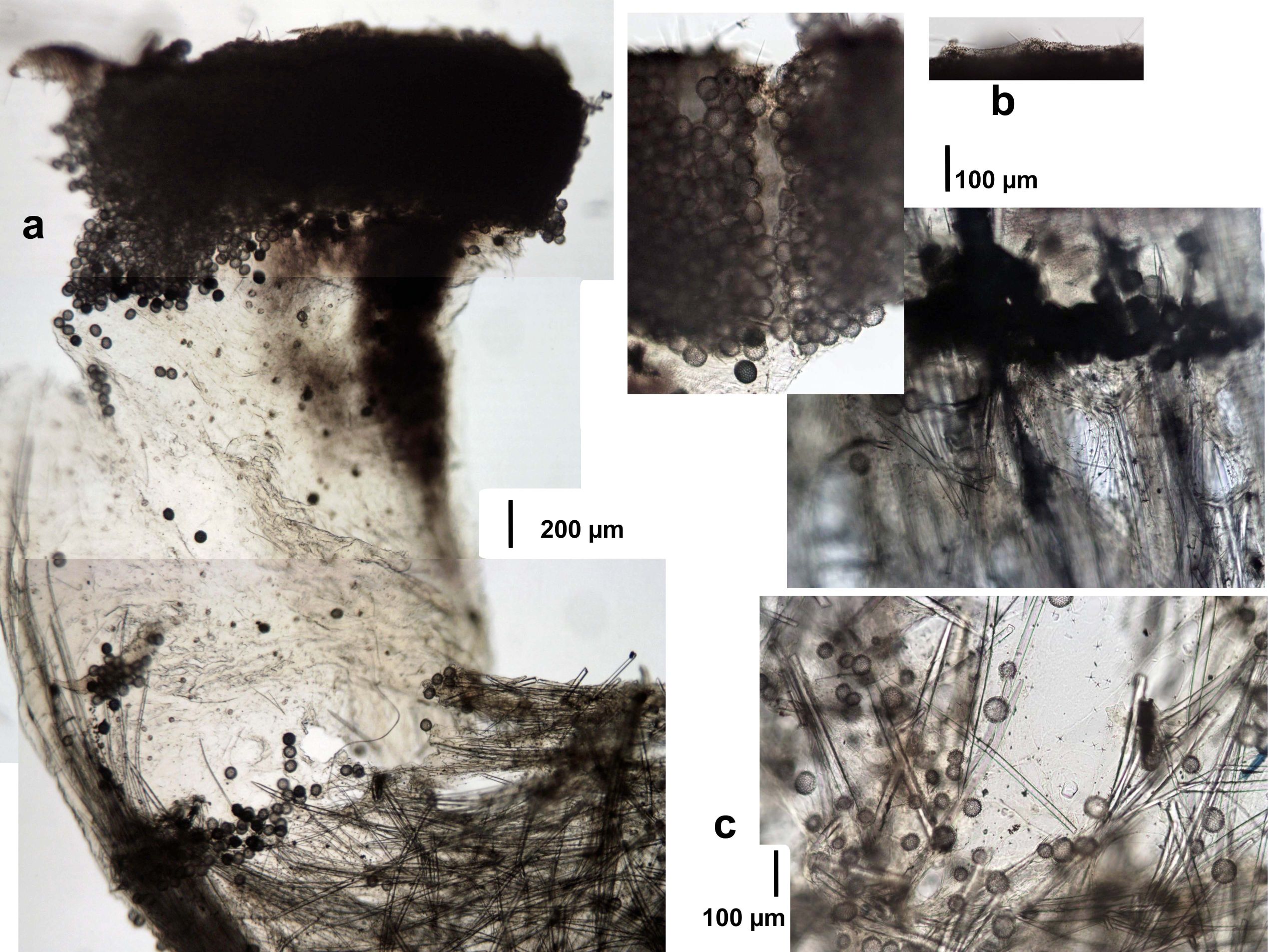
Tissue Image: a) Perpendicular section of the body tracts (surface above; the section was twisted upon drying); b) magnification of the surface (above), subsurface (mid) and choanosome (lower). Sample from the Bahamas.