
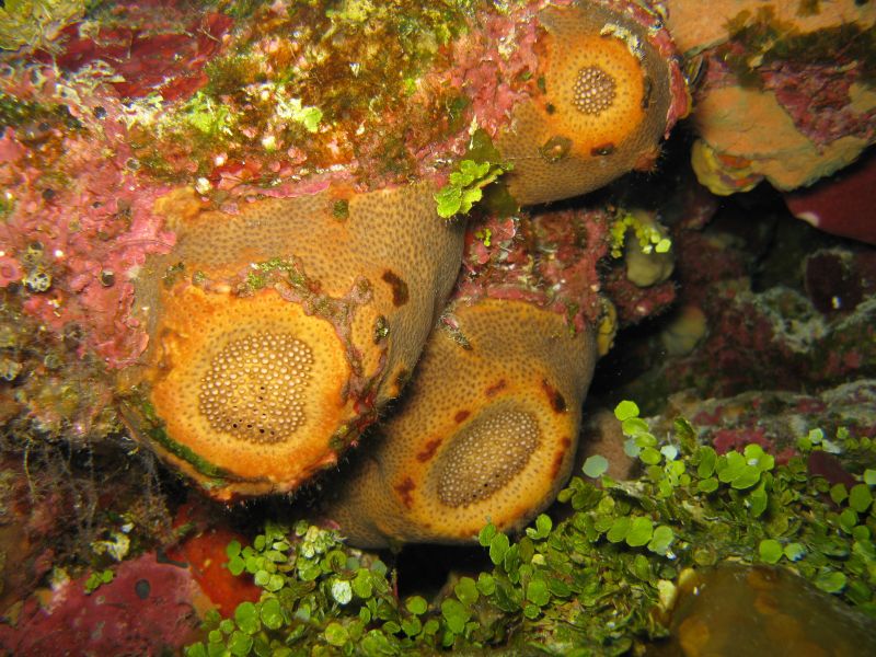
Species: Geodia aff. gibberosa sp.5
- Location: Little Inagua, Bahamas
- Habitat: deep reef
- Photographer: Sven Zea
- Picture Taken On: 2011-07-28 17:01:00
- Picture Notes: no image notes
- Color: cinnamon-tan, orange
- Morphology: massive
- Consistency: hard

Spicules Image: a) Oxeas-styloids-styles-strongyles I; b) anysostrongyloxeas-oxeas II, c) enlarged tips of oxeas-styloids-styles-strongyles I; d) enlargement of an anysostrongyloxea II; e) plagiotriaenes; f) microscleres (sterrasters, developed and developing); g) enlargement of microscleres (rossettes of sterrasters on the left; spined asters in 2-3 types, oxyasters and strongyl- or oxyasters). Sample from the Bahamas.
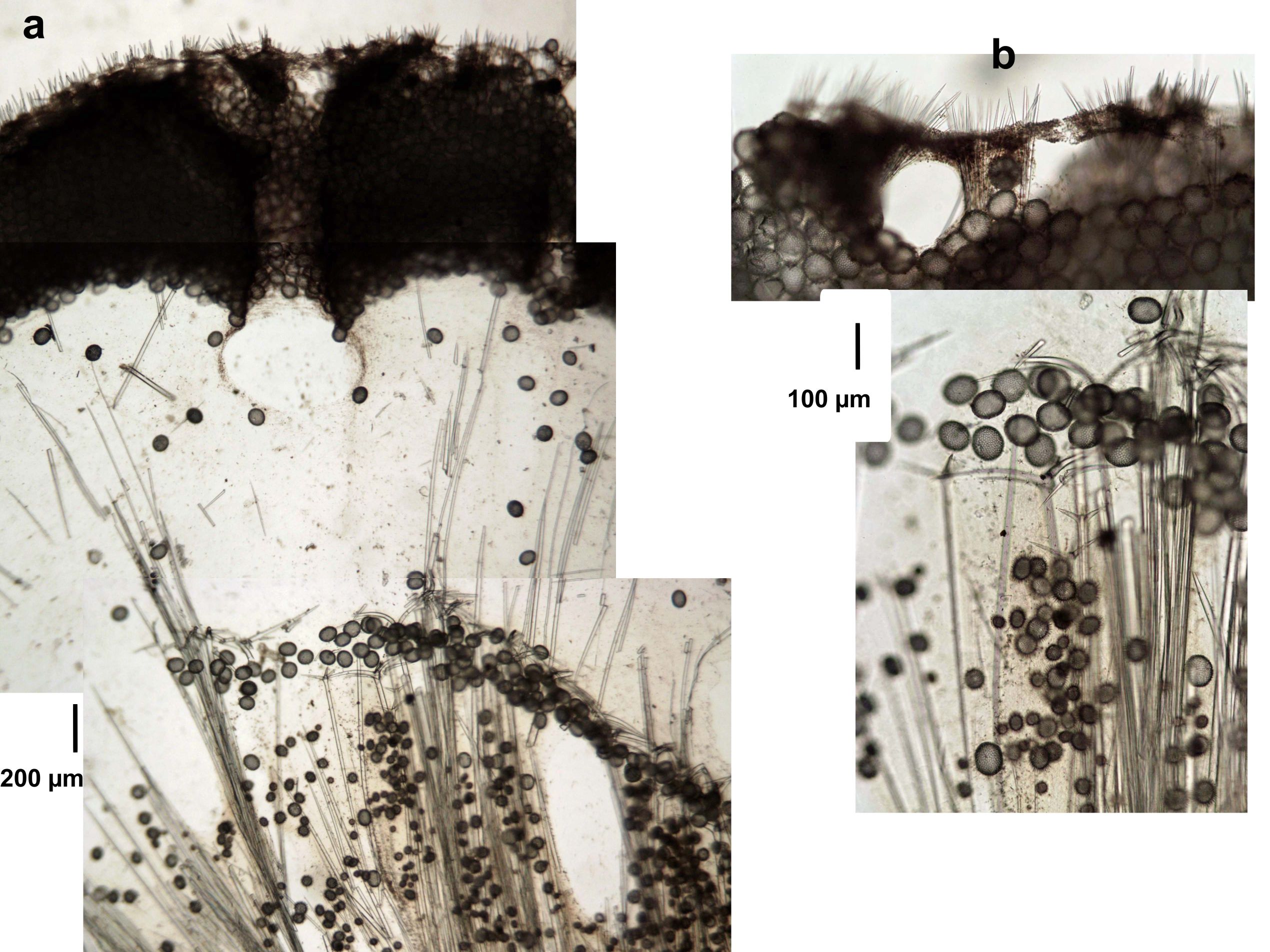
Tissue Image: a) Perpendicular section of the body (surface cortex above); b) magnification of the surface (above) and the subsurface (below). Sample from the Bahamas.