
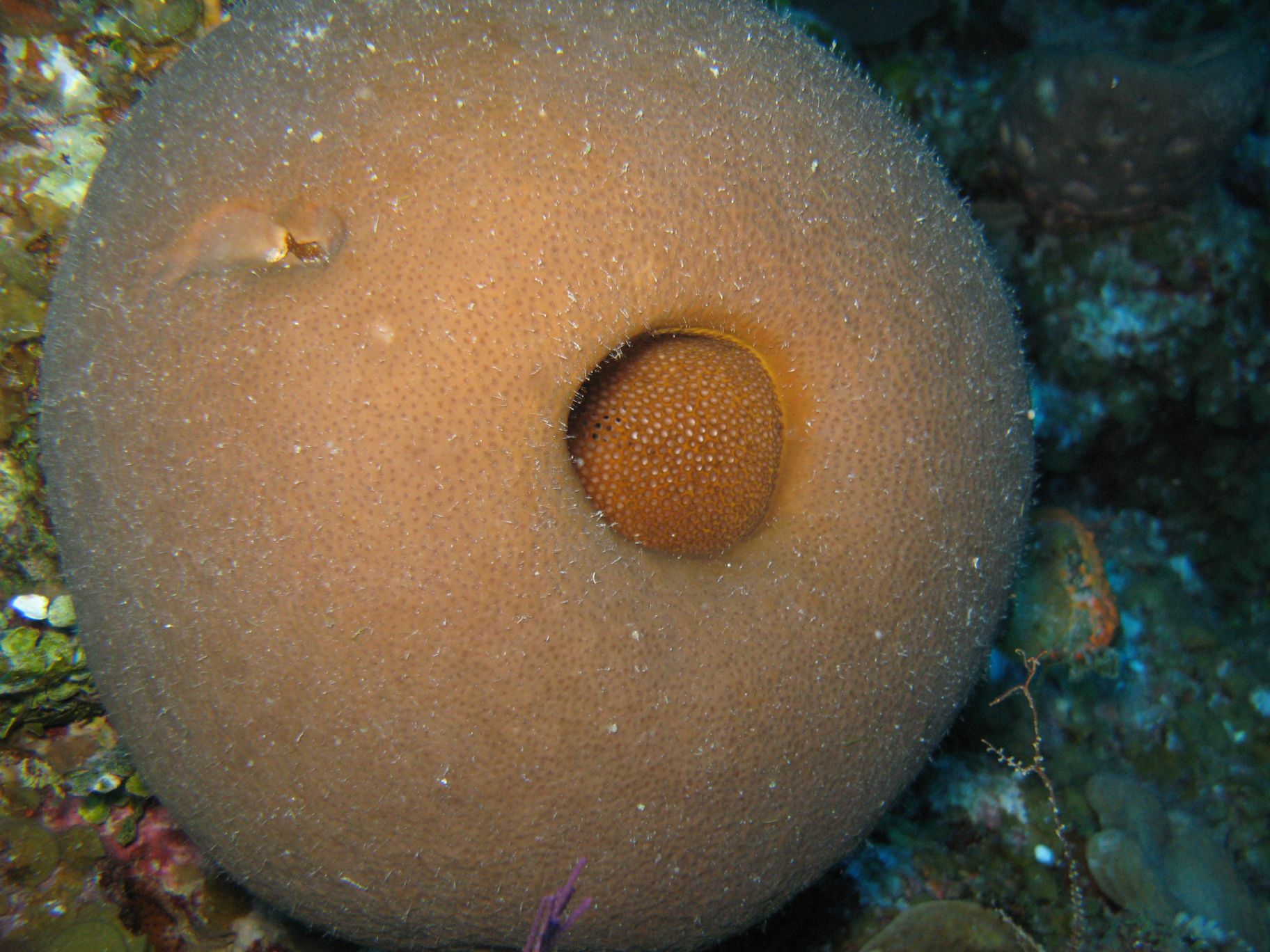
Species: Geodia aff. gibberosa sp.4-“melon”
- Location: Plana Cays, Bahamas
- Habitat: deep reef
- Photographer: Tse-Lyn Loh
- Picture Taken On: 2011-07-21 09:29:24
- Picture Notes: no image notes
- Color: cinnamon-tan
- Morphology: spherical
- Consistency: hard
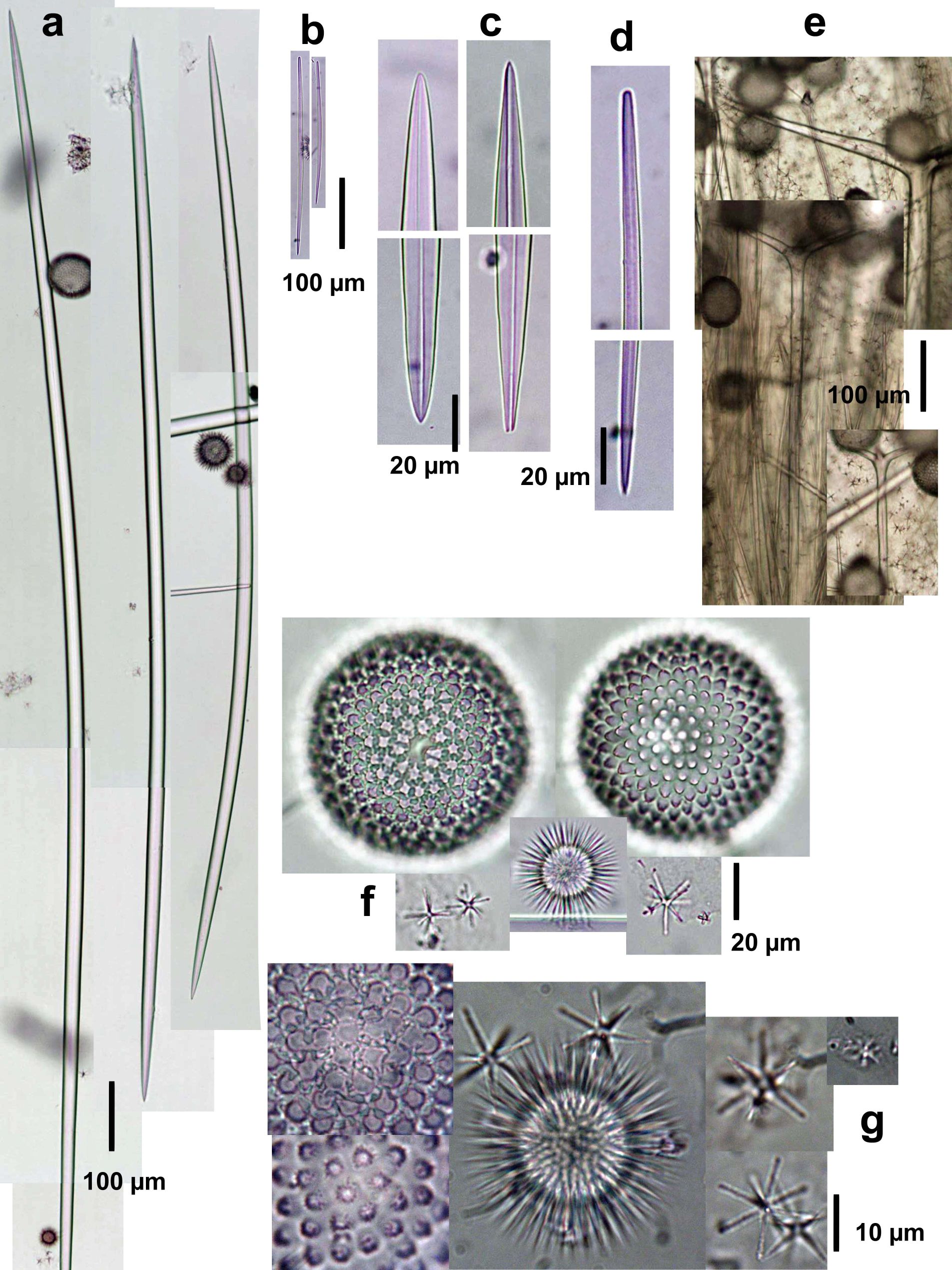
Spicules Image: a) Oxeas I; b) anysostrongyloxeas II, c) enlarged ends of oxeas I; d) enlargement of an anysostrongyloxea II; e) plagiotriaenes; f) microscleres (sterrasters, developed and developing; spined asters in two types, oxyasters and strongyl- or oxyasters); g) enlargement of microscleres (rossettes of sterrasters on the left). Sample from the Bahamas.
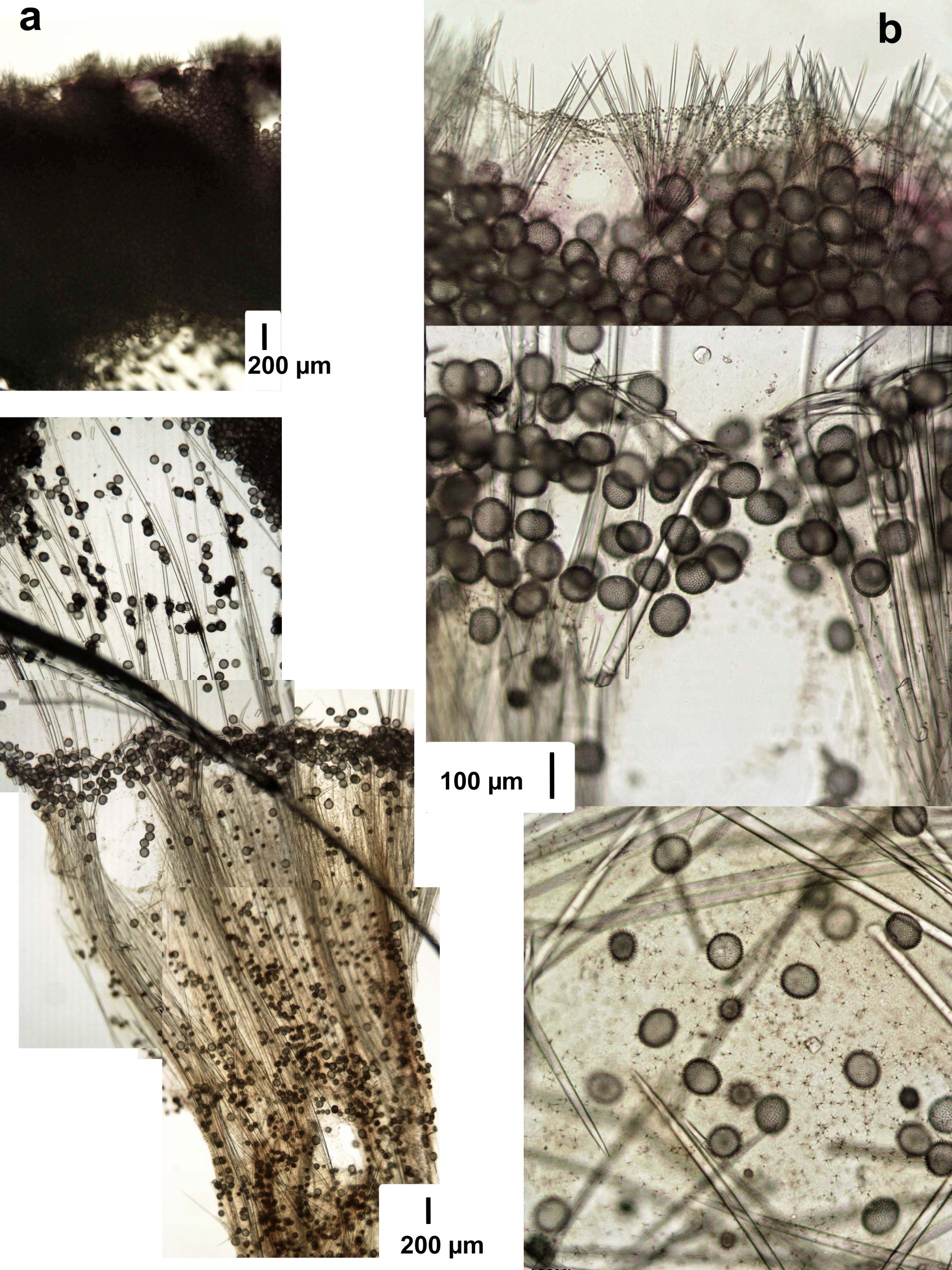
Tissue Image: a) Perpendicular section of the body (surface above; the dark curved line crossing the lower panel is a fracture on the slide cover slip glass); b) magnification at the surface (above), subsurface (mid) and choanosome (lower) showing sterrasters and the aligned cladomes of triaenes; the lower panel is a magnification of the choanosome. Sample from the Bahamas.