
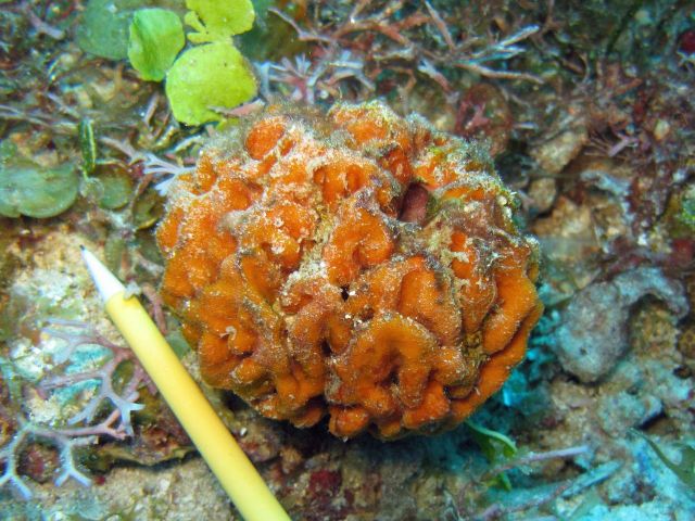
Species: Geodia aff. corticostylifera
- Location: Martinique
- Habitat: deep reef
- Photographer: Sven Zea
- Picture Taken On: 2013-12-06 00:00:00
- Picture Notes: no image notes
- Color: orange
- Morphology: bushy, spherical
- Consistency: tough
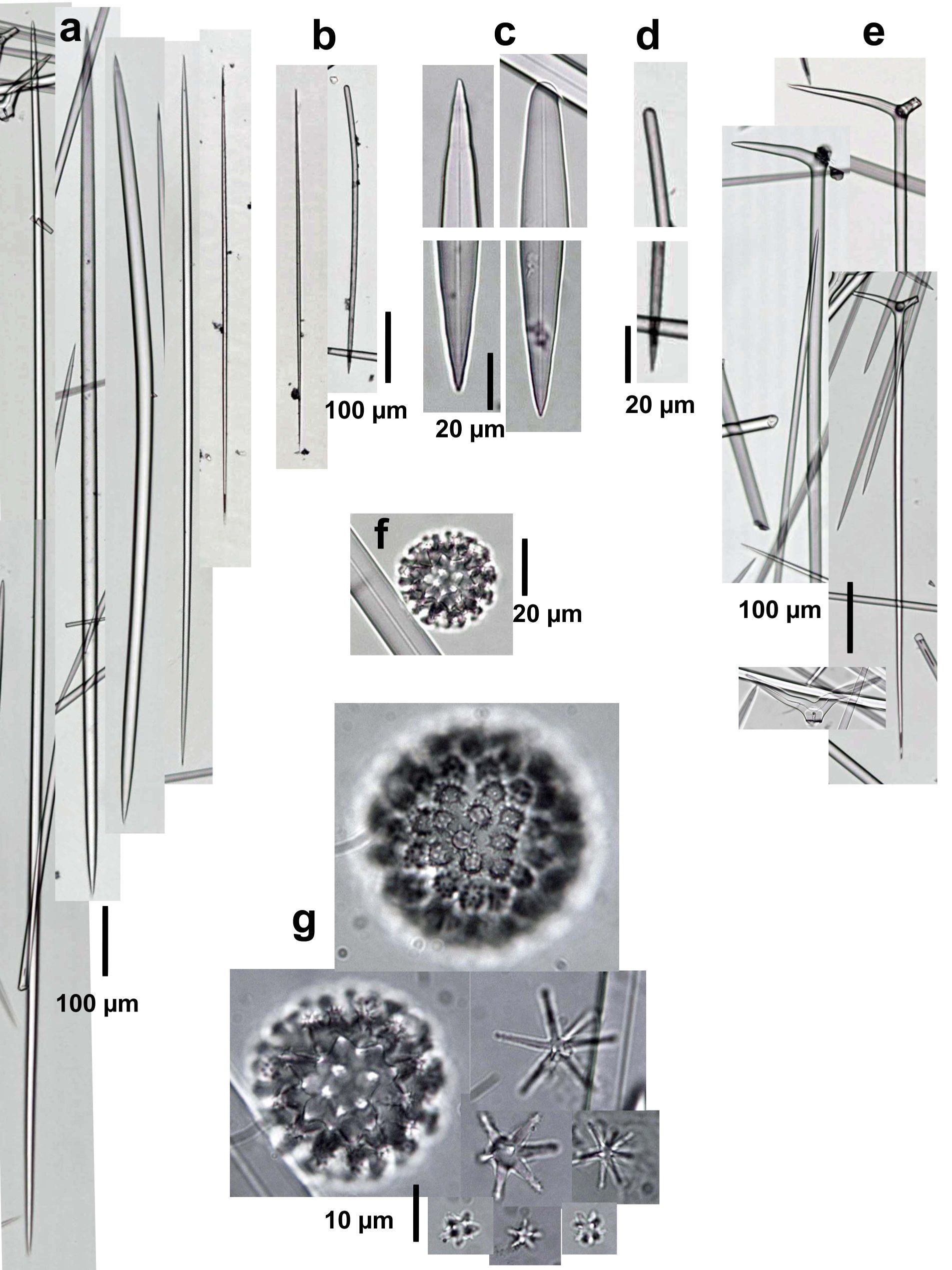
Spicules Image: a) Oxeas-anysostrongyloxeas I; b) oxeas-styles II, c) enlarged ends of oxeas-anysostrongyloxeas I; d) enlargement of a style II; e) plagiotriaenes; f) microscleres (sterraster); g) enlargement of microscleres (sterrasters, developing and fully developed; spined asters in 3 types, oxyasters and strongyl- or oxyasters). Sample from Martinique.
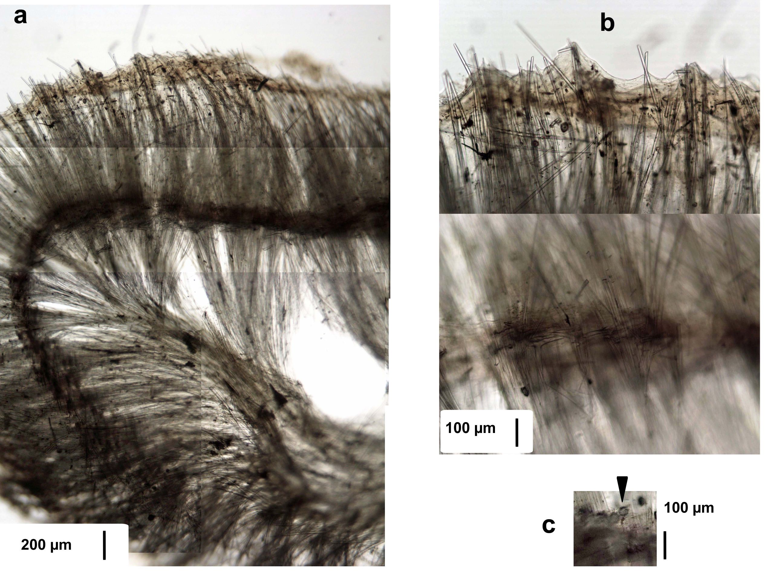
Tissue Image: a) Perpendicular section at a a surface fold; b) magnification at the surface (above) and subsurface (below); c) magnification the subsurface showing a few sterrasters (arrow). Sample from Martinique.