
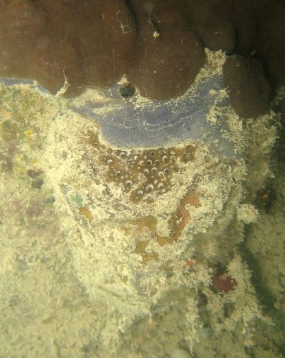
Species: Geodia sp.-“Biscayne Bay”
- Location: Florida, Biscayne Bay, United States
- Habitat: mangrove/coastal lagoon
- Photographer: Sven Zea
- Picture Taken On: 2010-07-01 16:00:02
- Picture Notes: The blue strip on top of the sponge is an unidentified sponge of the family Chalinidae (Haplosclerida). The dark brown lobes on top are the colonial tunicate Eudistoma hepaticum.
- Color: brown
- Morphology: massive
- Consistency: tough
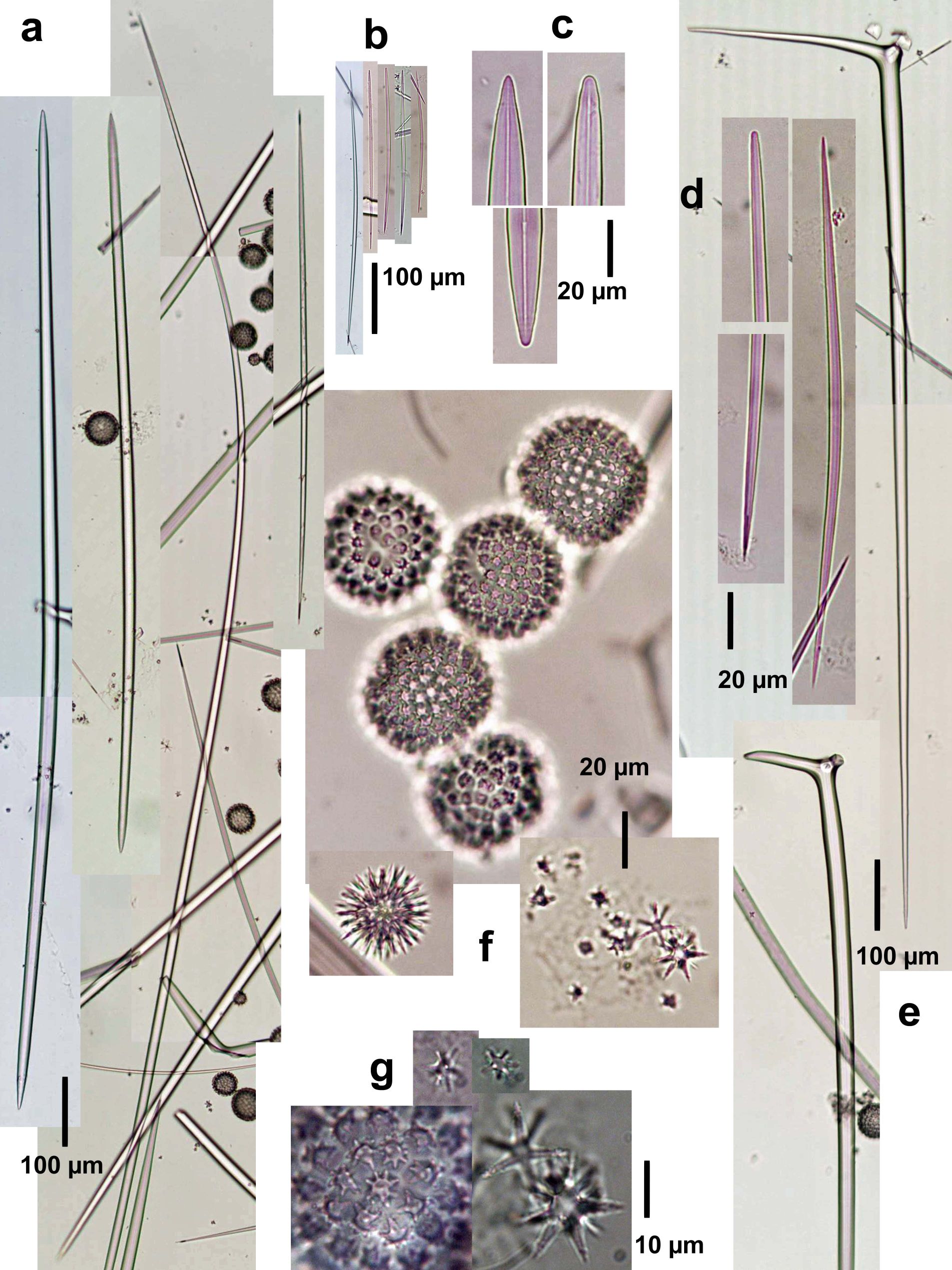
Spicules Image: a) Oxeas I; b) anysostrongyloxeas-oxeas II, c) enlarged end of oxeas I; d) enlargement of oxeas-anysostrongyloxeas II; e) plagiotriaenes; f) microscleres (sterrasters, developed and developing; spined asters in 2-3 types, oxyasters and strongyl- or oxyasters); g) enlargement of microscleres (rossettes of sterrasters on the left). Sample from South Florida.
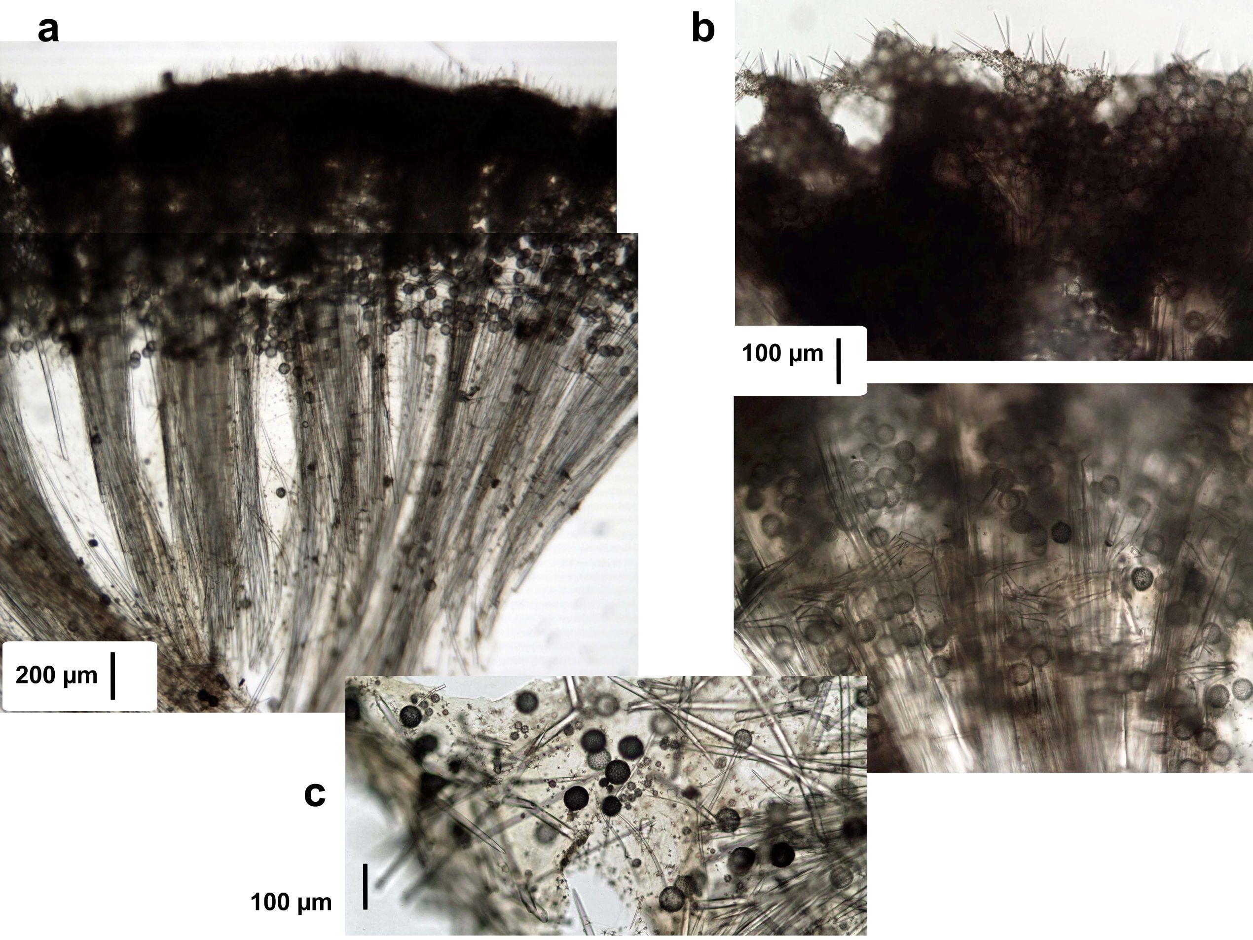
Tissue Image: a) Perpendicular section at the surface; b) magnification at the surface (above) and subsurface (below); c) magnification of the choanosome. Sample from South Florida.