
- Location: Colombia-Santa Marta
- Habitat: rocky shore/shallow reef
- Photographer: Sven Zea
- Picture Taken On: 1983-04-28
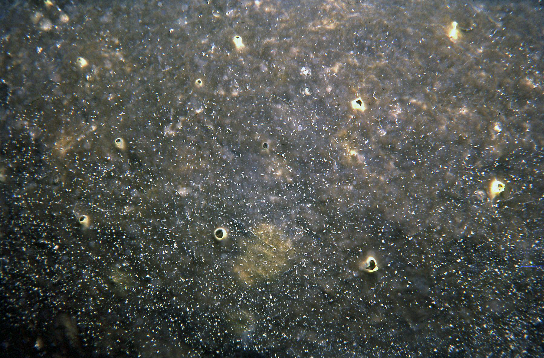
- Picture Notes: live photograph of the holotype ICN-MHN(Po) 0226
- Color: brown
- Morphology: encrusting
- Consistency: tough
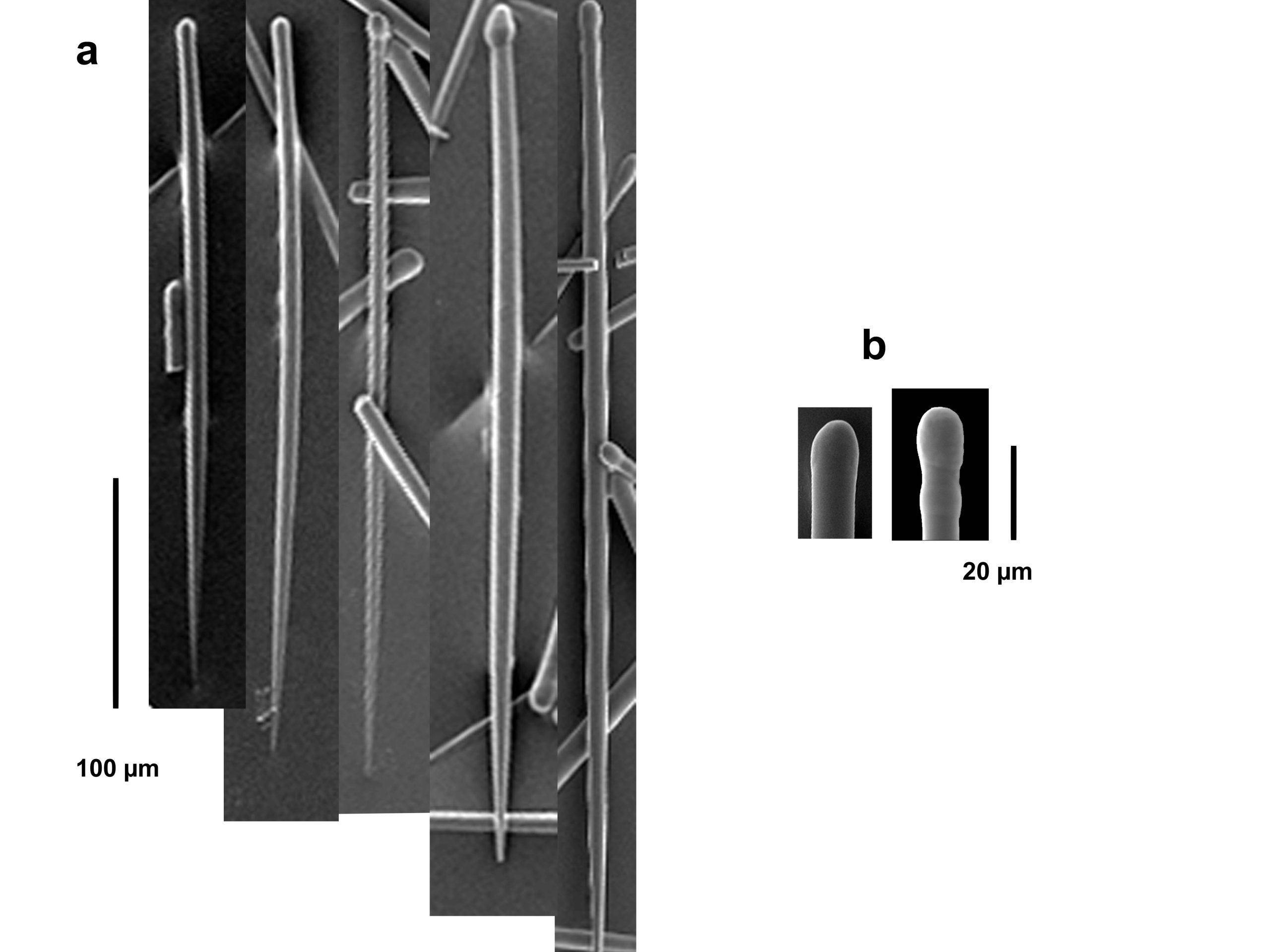
Spicules image: SEM images of: a) Tylostyles; b) ends of tylostyles (SEM). Sample from Santa Marta, Colombia (specimen ICN-MHN(Po) 0226, holotype).
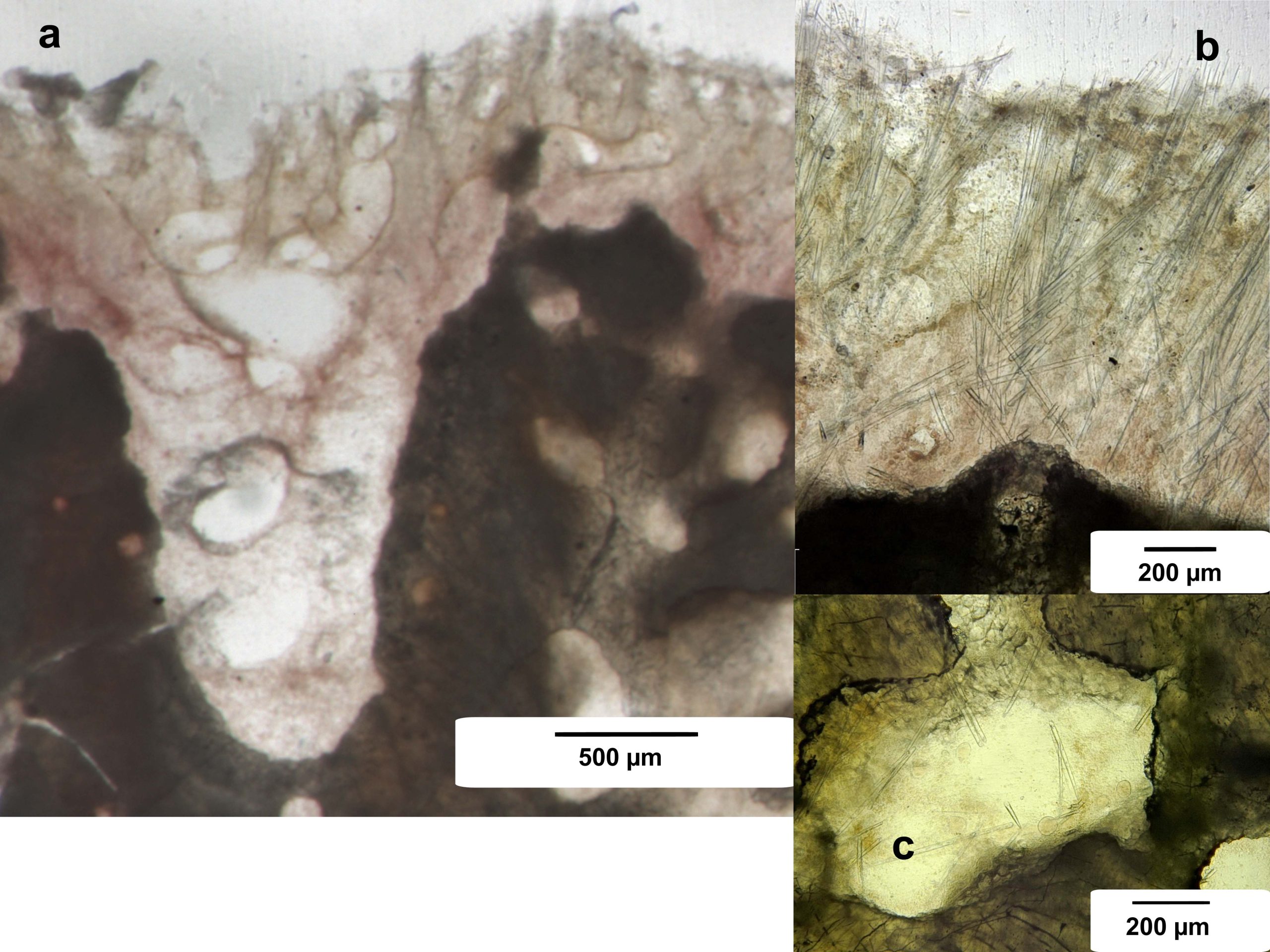
Tissue image: Ground and polished sections of coral skeleton with sponge: a) perpendicular section at the surface showing the epilithic portion of the tissue over excavated areas; b) perpendicular section at the surface showing brushes of spicules and inhalant canals; c) enlargement of the surface palisade; d) enlargement of an excavated portion of the coral skeleton showing the characteristic etch marks. Sample from Santa Marta, Colombia (specimen ICN-MHN(Po) 0226, holotype).