
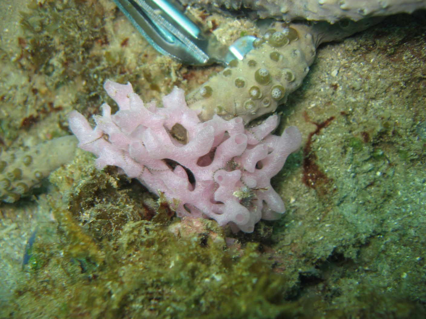
- Location: Bocas del Toro, Panama
- Habitat: deep reef
- Photographer: Cristina Diaz
- Picture Taken On: 2010-07-25 00:00:00
- Picture Notes: no image notes
- Color: pink-lilac
- Morphology: tube
- Consistency: crumbly
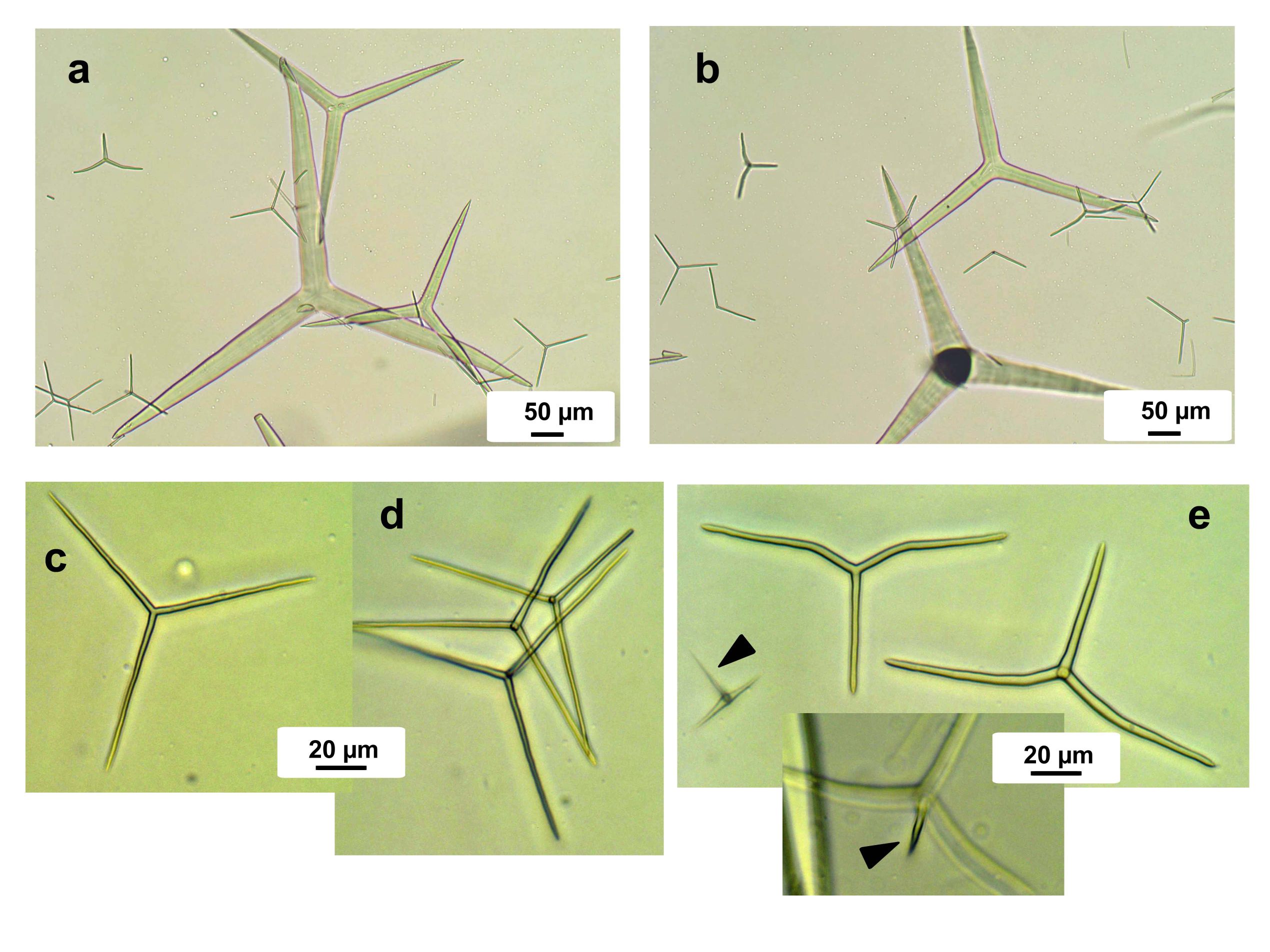
Spicules Image: a) And b) large tri and tetractines II (cortical) and smaller tri and tetractines I, normal and sagittal (choanosomal); c) and d) tri- an tetractines I (choanosomal); e) sagittal tri and tetractines, showing the apical actine of a tetractine (arrow in inset). Photos courtesy of Michelle Klautau. Sample from the Bocas del Toro, Panama, by Cristina Diaz.
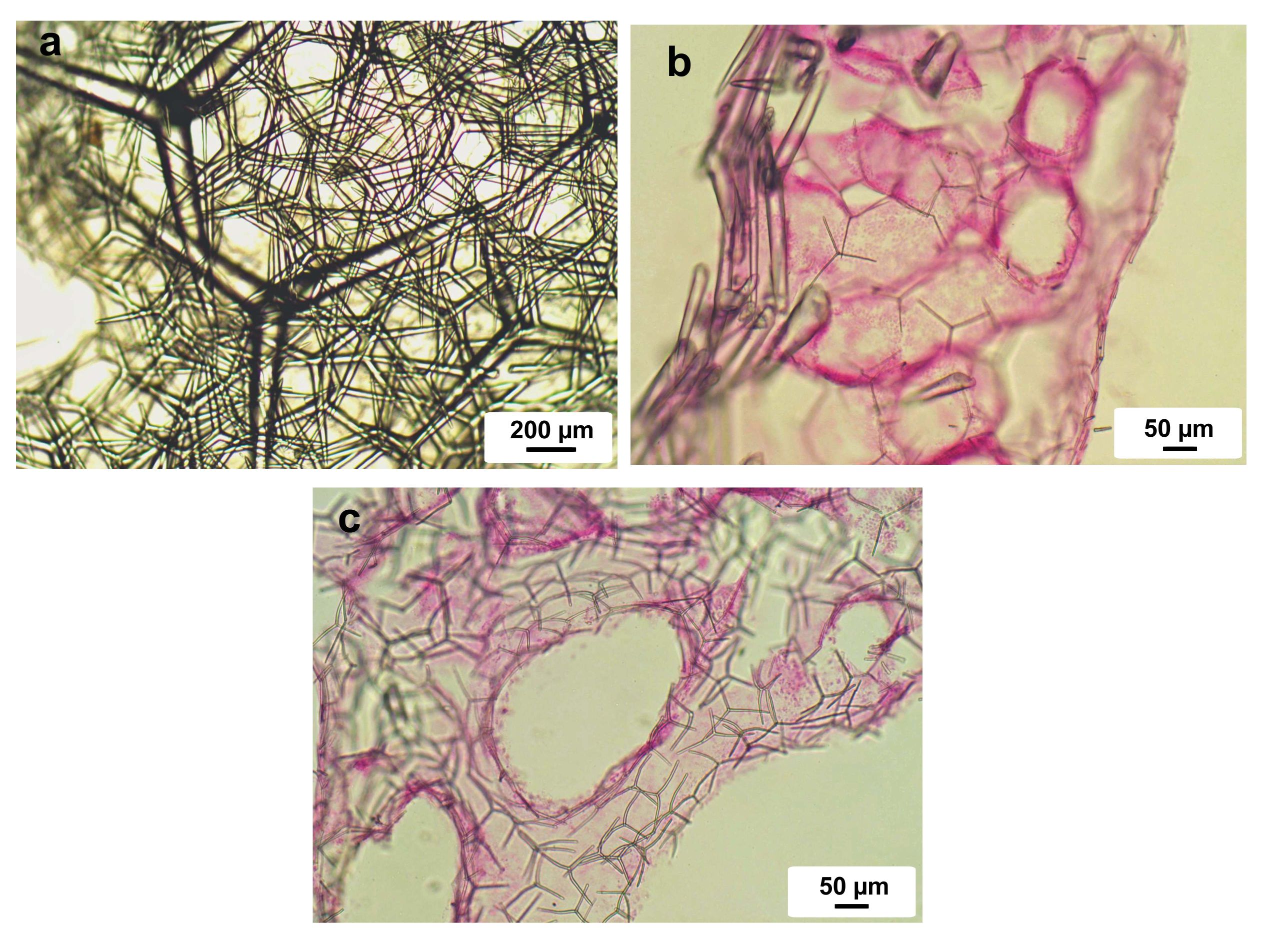
Tissue Image: a) Top view of the surface cortex; b) histological cross section of a tube; c) choanosome. Sample from Bocas del Toro, Panama. Photos courtesy of Michelle Klautau.