
Species: Myrmekioderma gyroderma
- Location: Little San Salvador, Bahamas
- Habitat: deep reef
- Photographer: Sven Zea
- Picture Taken On: 2004-06-21 09:03:56
- Picture Notes: no image notes
- Color: orange-yellow
- Morphology: massive
- Consistency: crumbly

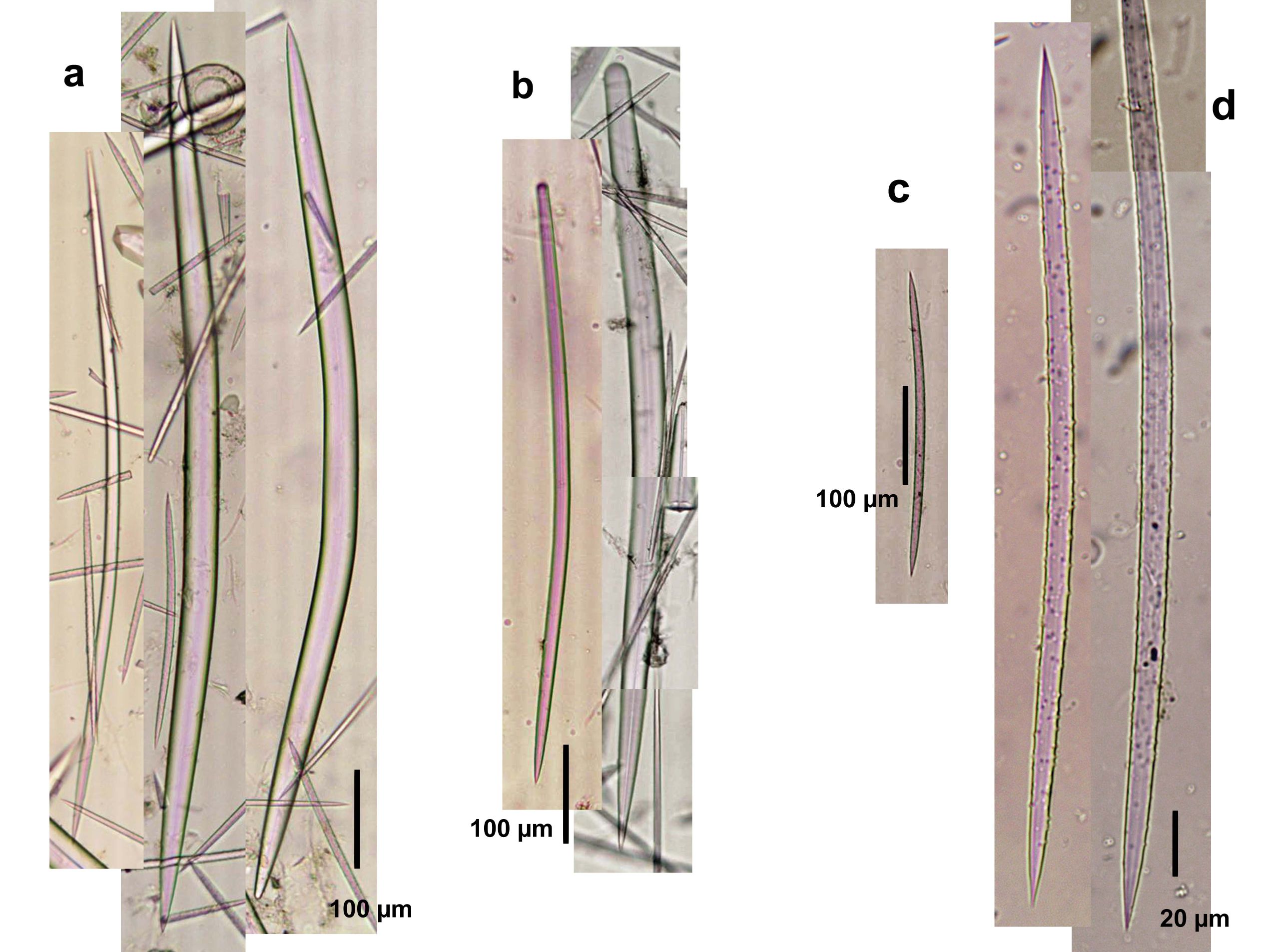
Spicule Images: a) Oxeas I; b) style modifications; c) oxea II (acanthoxea); d) magnification of oxea II. Sample from Santa Marta, Colombia.
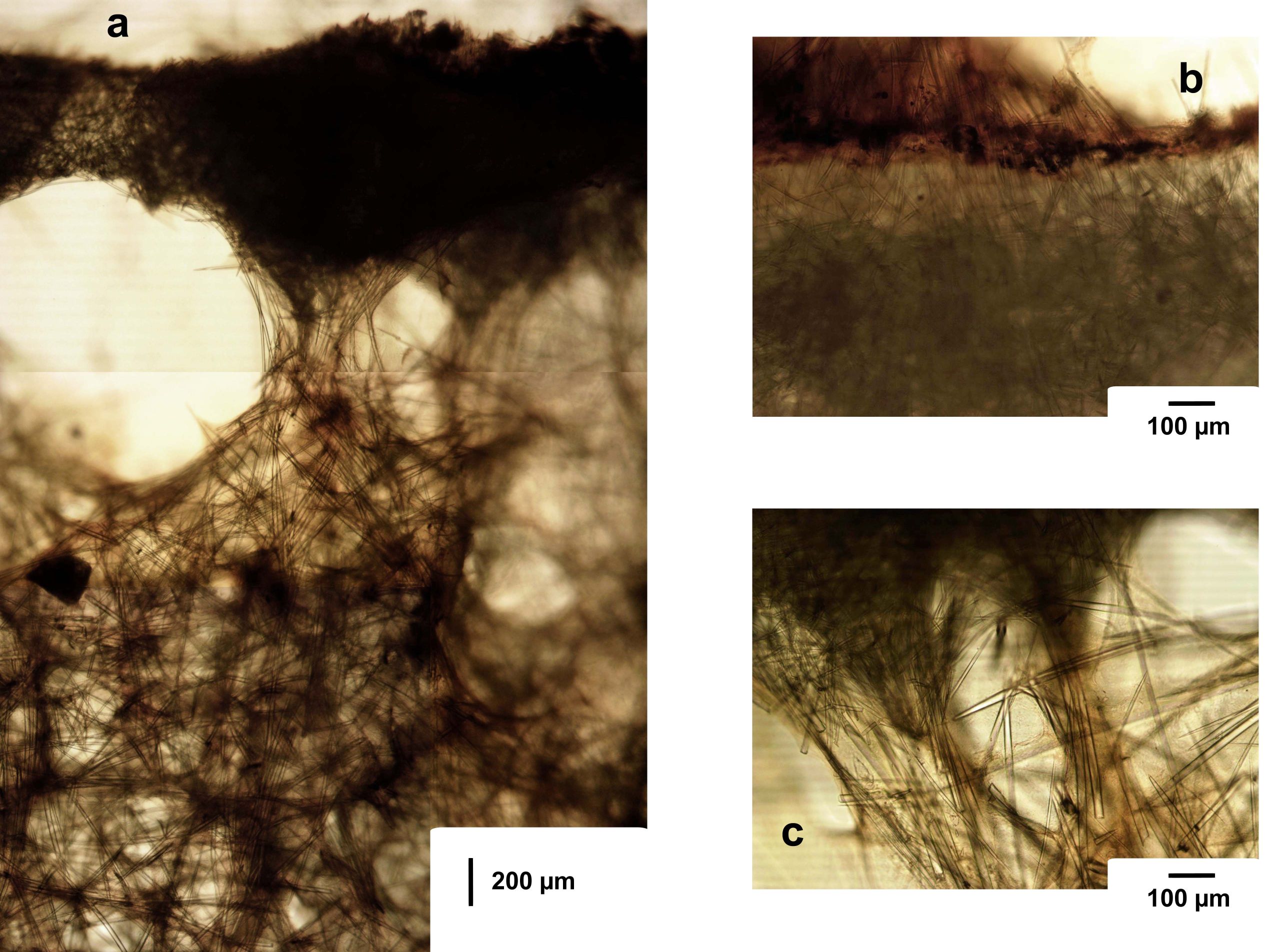
Tissue Images: a) Perpendicular section of the body; b) magnification of the cortex; c) magnification of a pillar supporting the cortex. Sample from the Bahamas.