
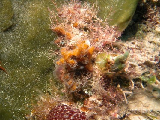
- Location: Sweetings Cay, Bahamas
- Habitat: deep reef
- Photographer: Sven Zea
- Picture Taken On: 2007-06-16 21:32:56
- Picture Notes: Orange bushy branchelets in the center. Green sponge on t he left is Petrosia weinbergi
- Color: orange
- Morphology: bushy
- Consistency: crumbly
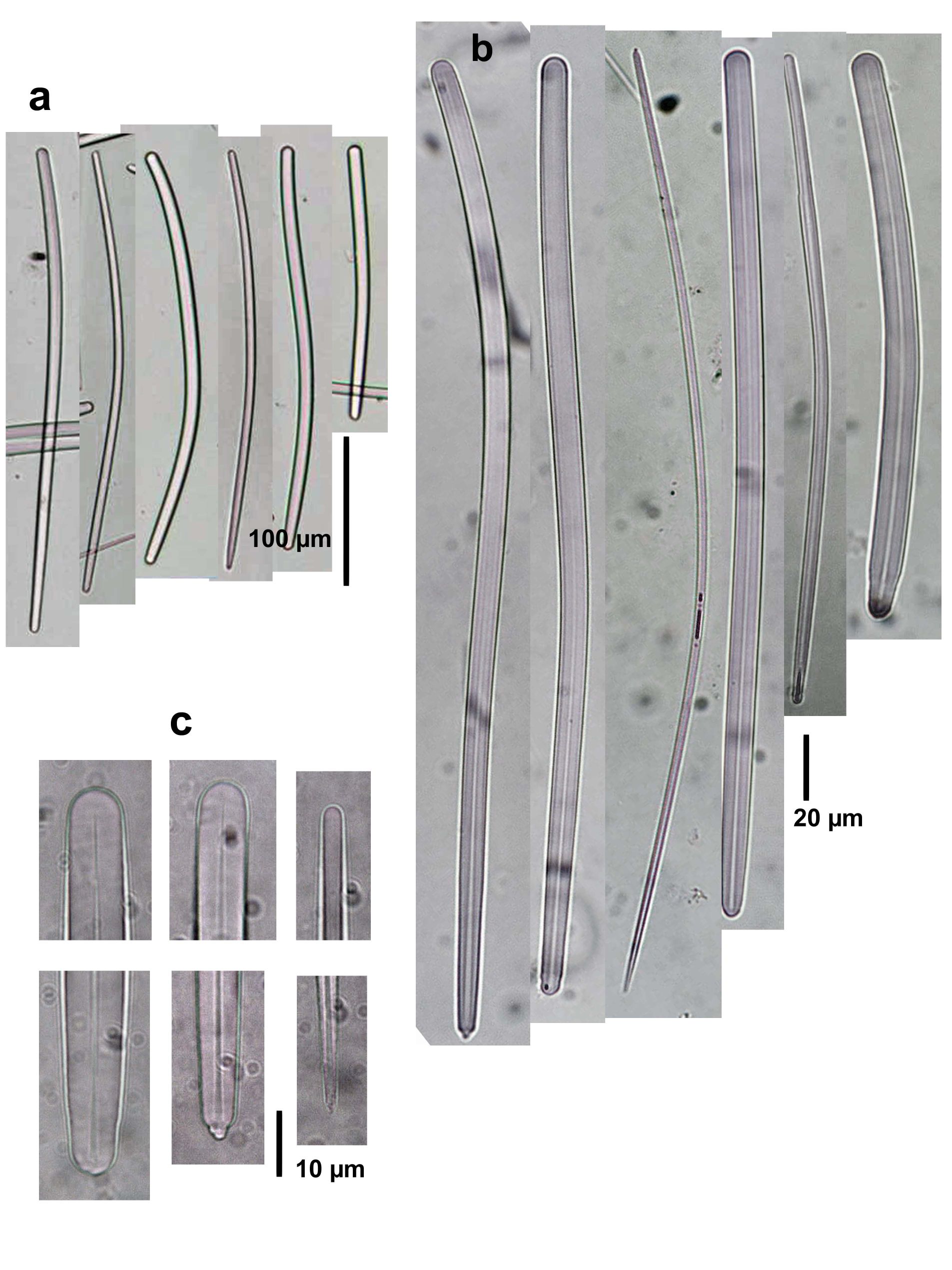
Spicule Images: a), b) Styloids at two different magnifications, with thinner, developmental stages tending to be styles; c) endings of spicules. Sample from the Bahamas.
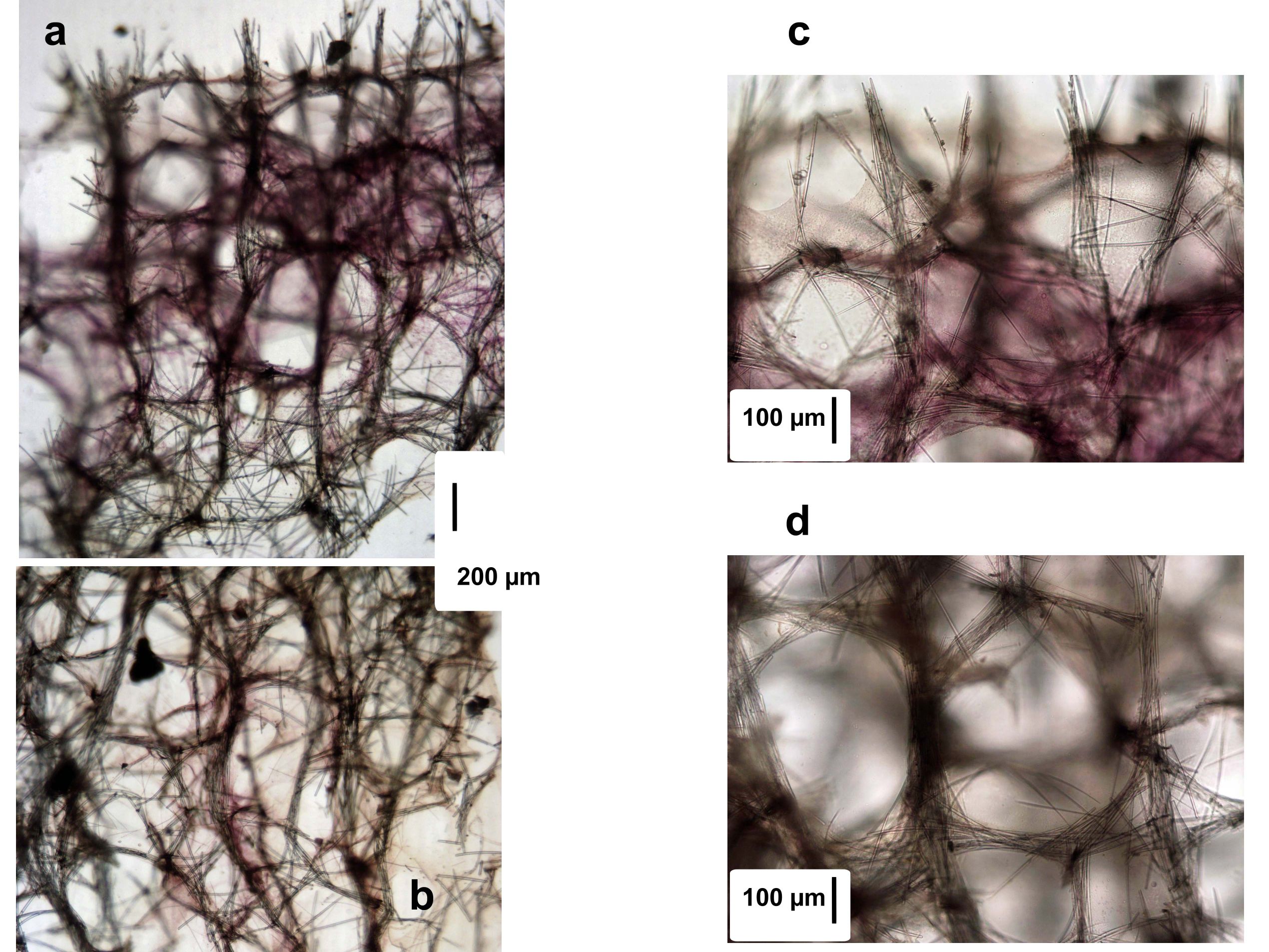
Tissue Images: a) Perpendicular section at the surface; b) view of the choanosome; c) view at the surface; c) view of the choanosome. Sample from the Bahamas.