
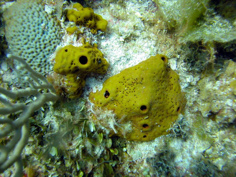
- Location: Little San Salvador, Bahamas
- Habitat: deep reef
- Photographer: Sven Zea
- Picture Taken On: 2004-06-21 14:43:25
- Picture Notes: no image notes
- Color: yellow
- Morphology: encrusting, massive
- Consistency: crumbly
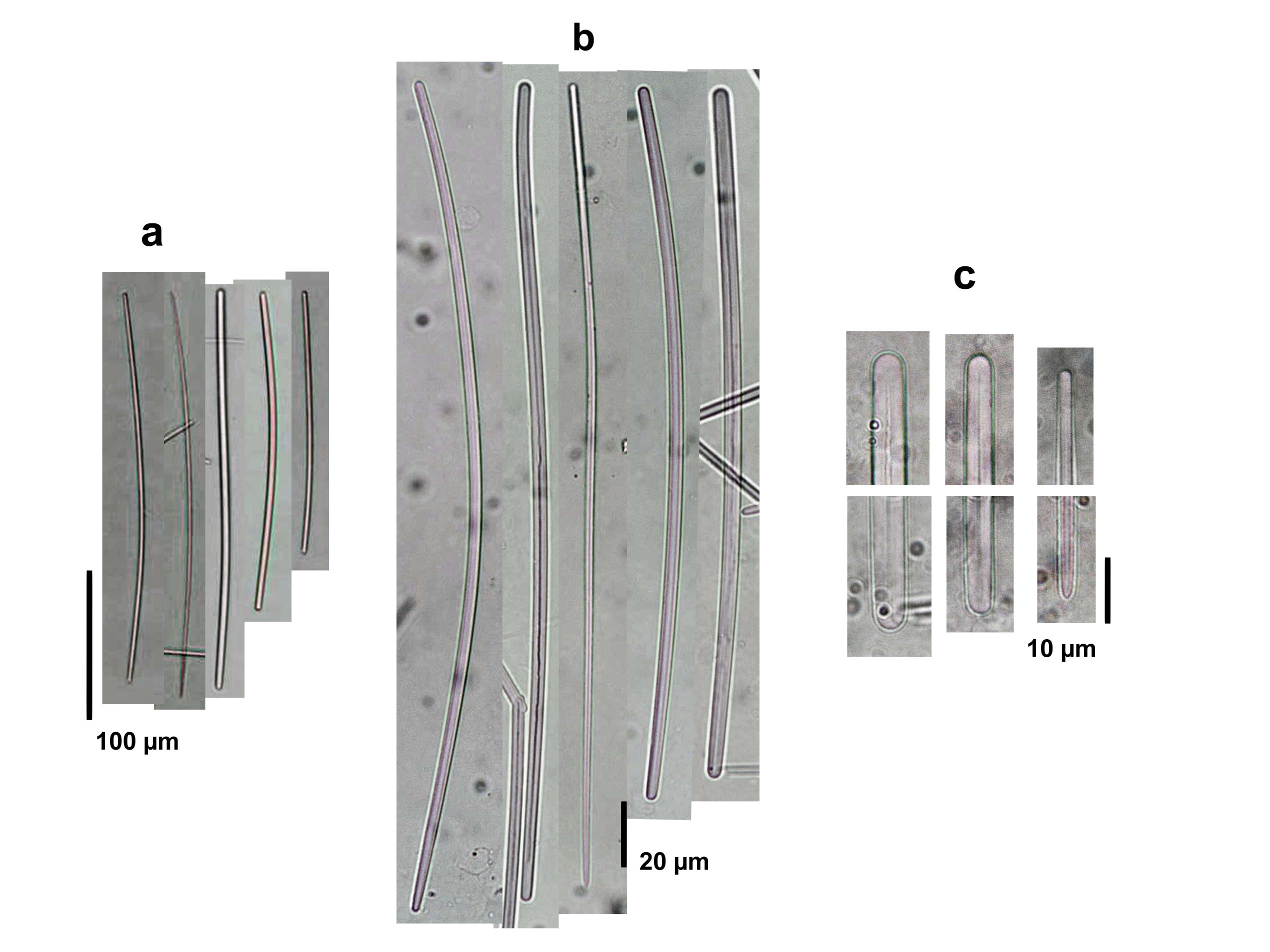
Spicule Images: a), b) Strongyles to styloids at two different magnifications, with thinner, developmental stages tending to be more styloid; c) endings of spicules. Sample from the Bahamas.
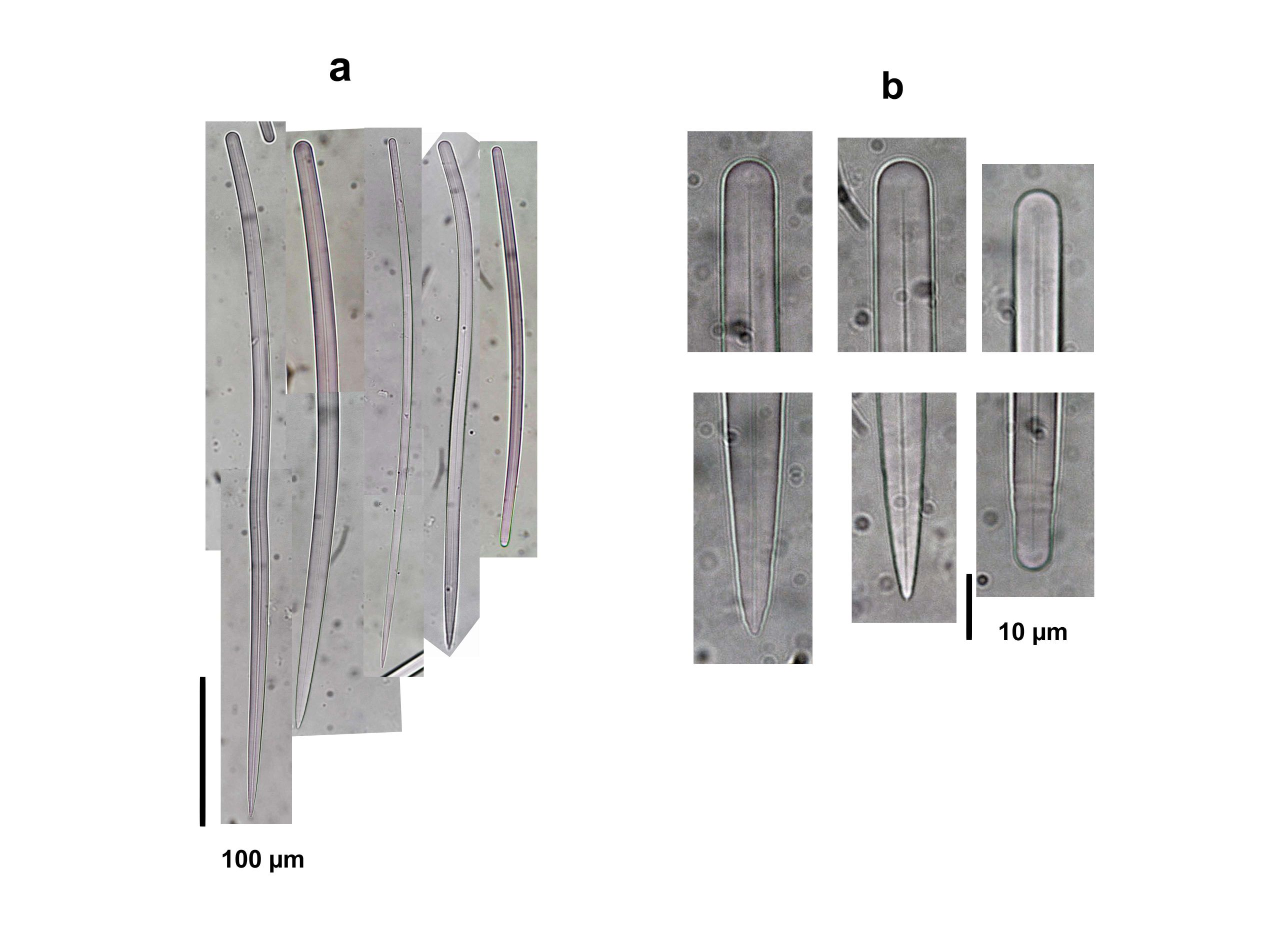
Spicule Images: a) Styles and styloids; b) endings of spicules. Sample from the Islas del Rosario, Colombia.
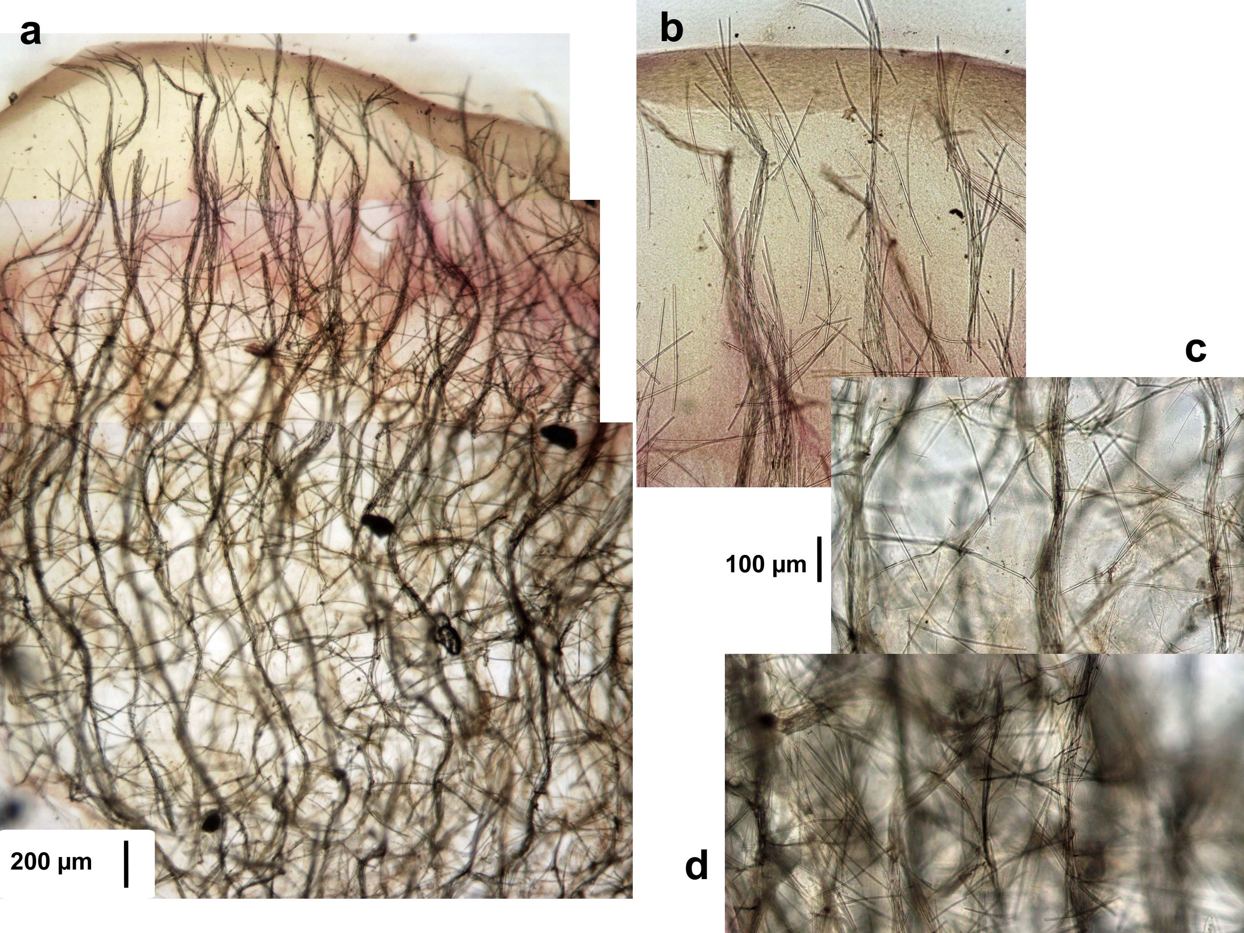
Tissue Images: a) Perpendicular section at the surface; b) view at the surface; c) view of the upper choanosome; d) view of the deeper choanosome. Sample from the Bahamas.