
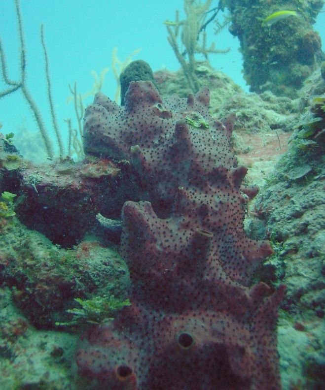
- Location: Little San Salvador, Bahamas
- Habitat: deep reef
- Photographer: Joseph Pawlik
- Picture Taken On: 2003-07-15 11:47:21
- Picture Notes: no image notes
- Color: brown
- Morphology: massive, tube
- Consistency: crumbly
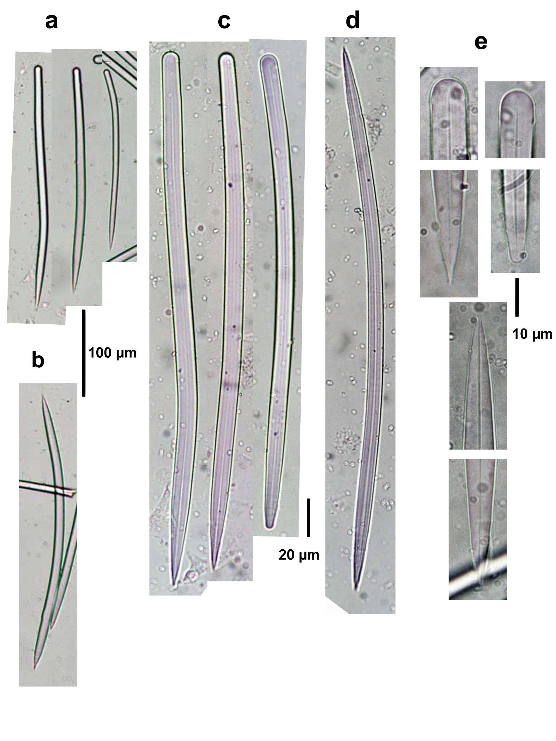
Spicule Images: a), c) Styles to styloids at two different magnifications; b), d) oxea at two different magnifications; e) ends of spicules. Sample from Islas del Rosario, Colombia.
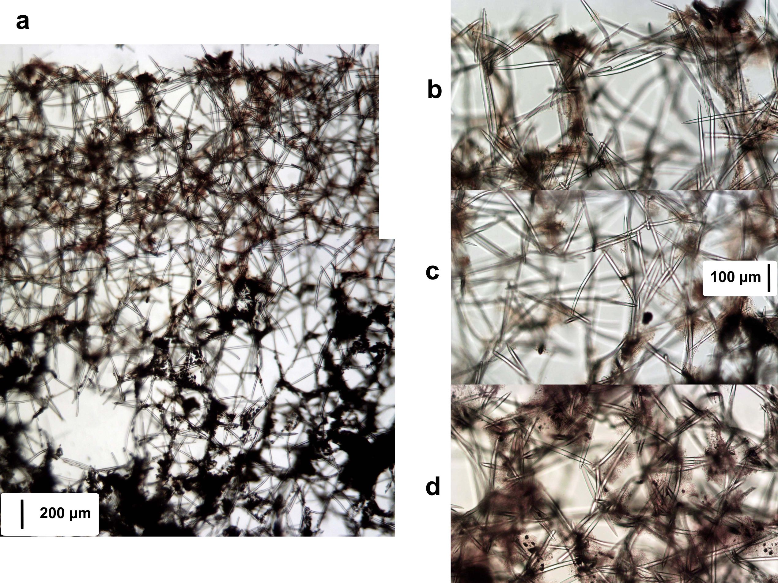
Tissue Images: a) Perpendicular section at the surface (dark granular cells appear to have been lost in the upper surface); b) view at the surface; c) enlargement of the choanosome; d) tangential view of the ectosome. Sample from Islas del Rosario, Colombia.