Observed Characteristics:
Color:
- brown
Morphology:
- encrusting
Consistency:
- tough
Locations:
- Colombia
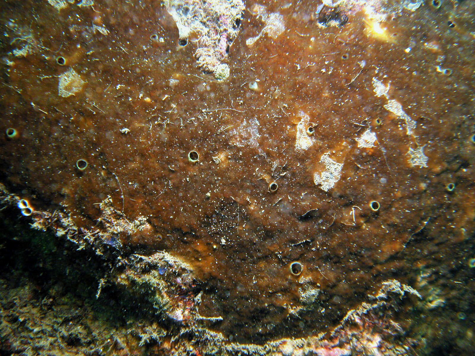
Species Description and Notes
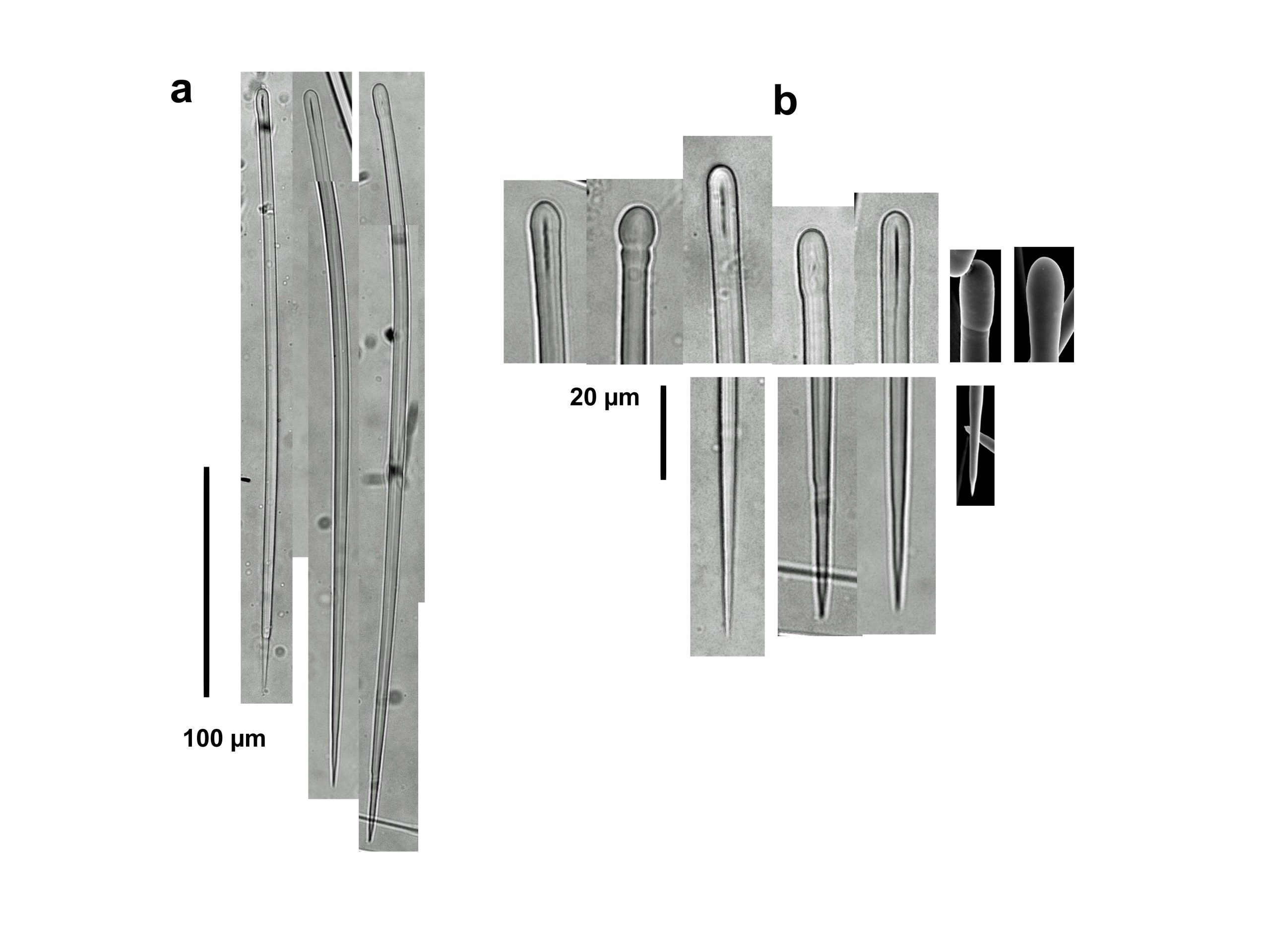
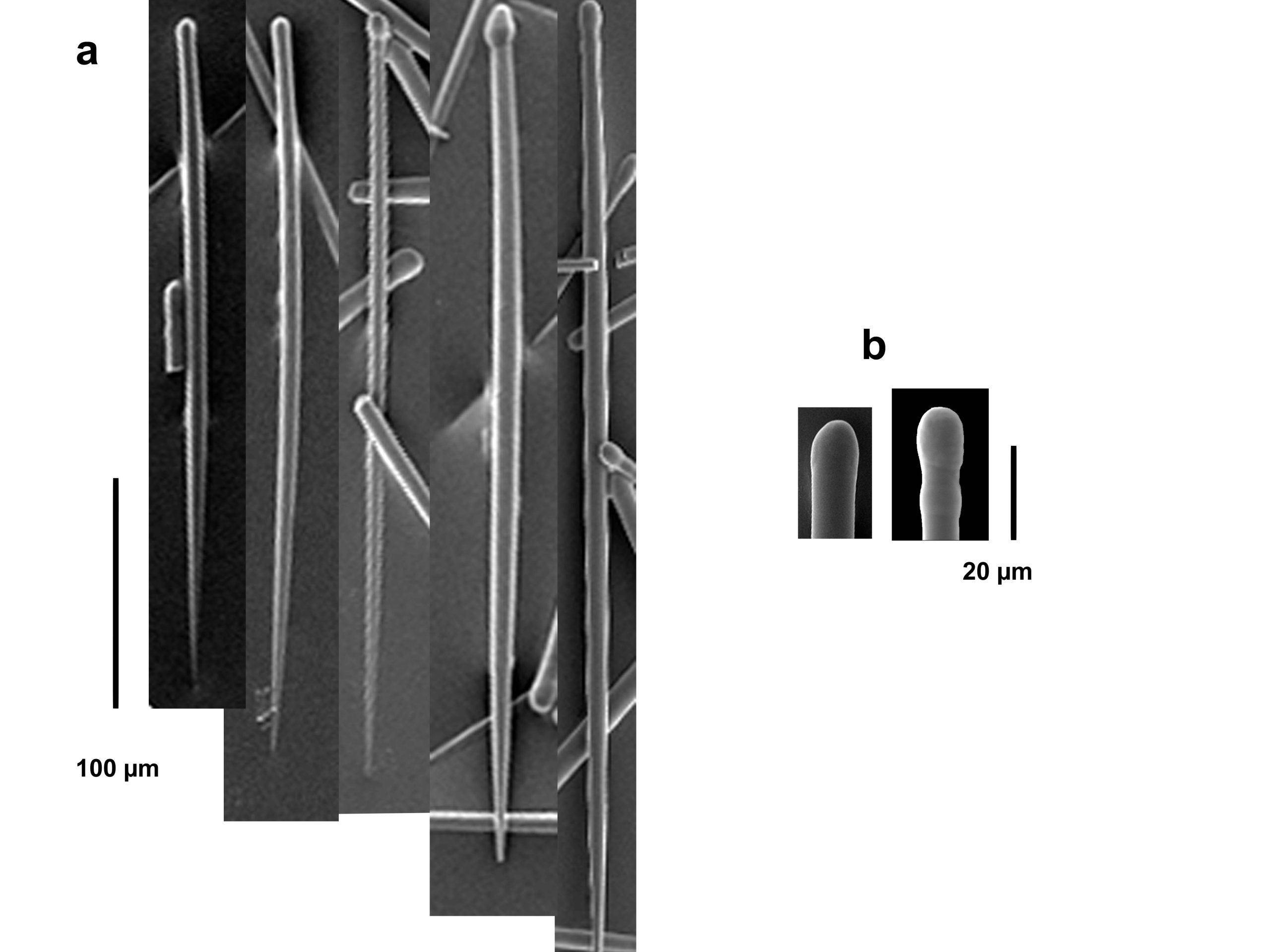
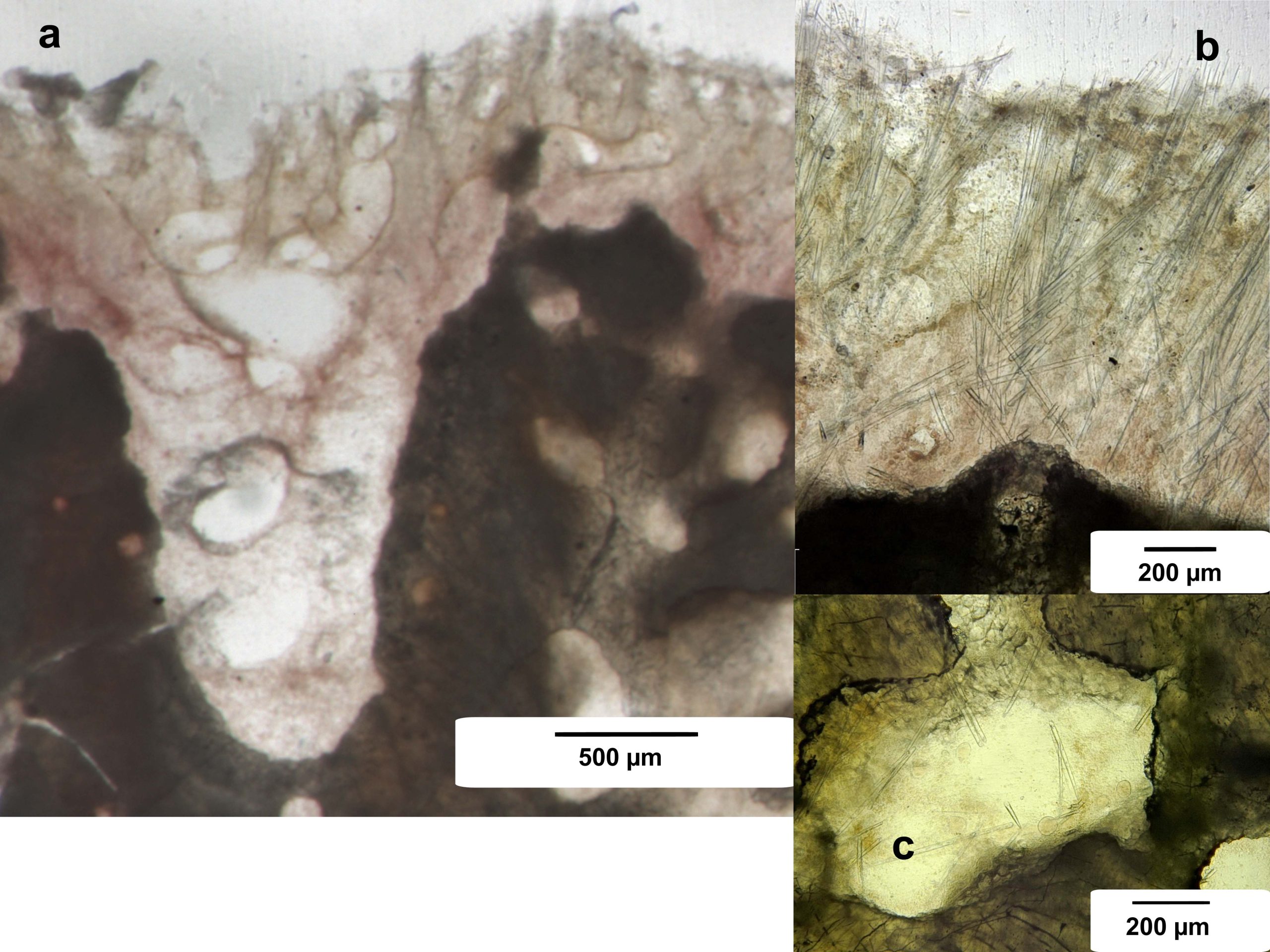
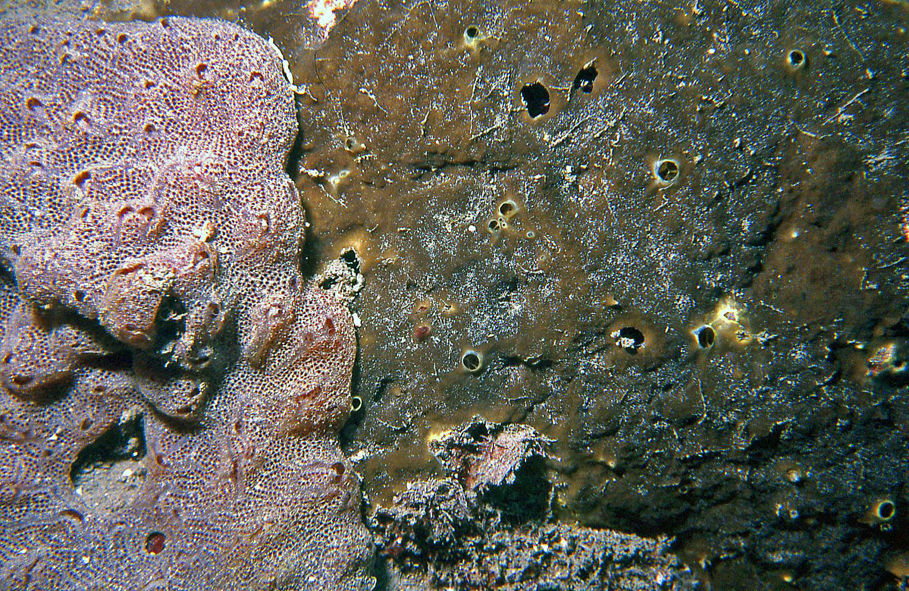
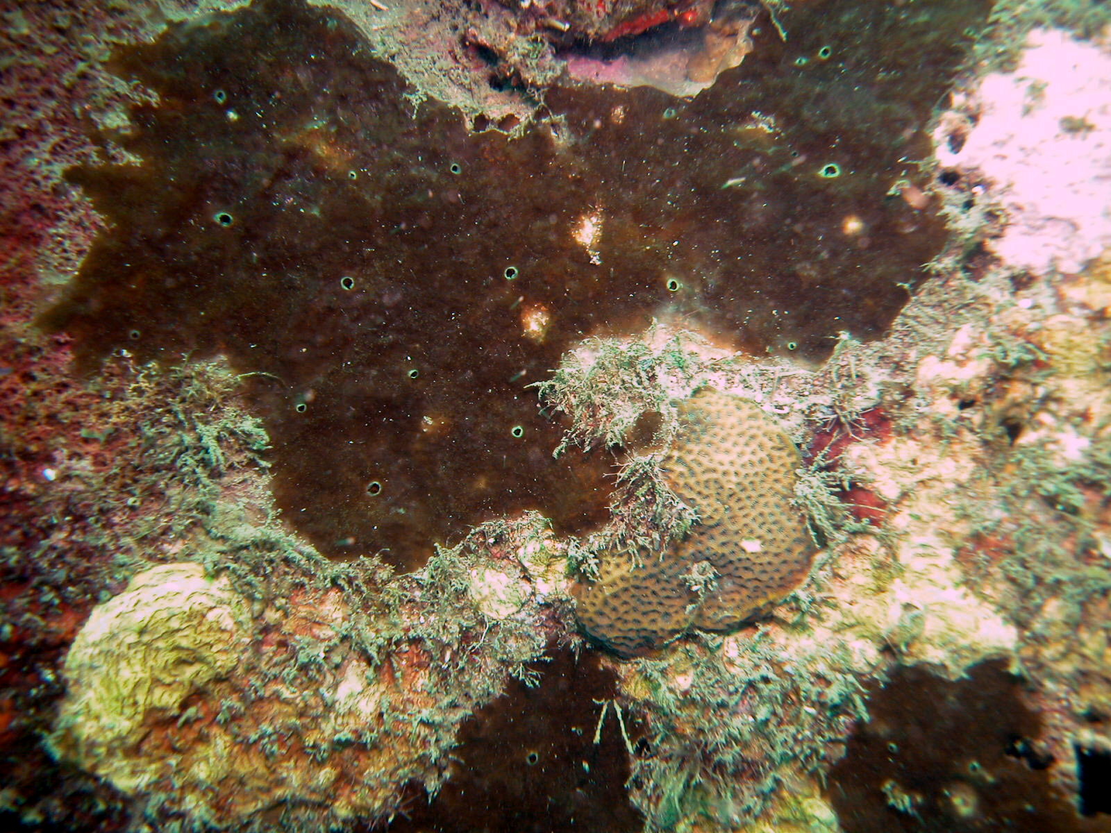
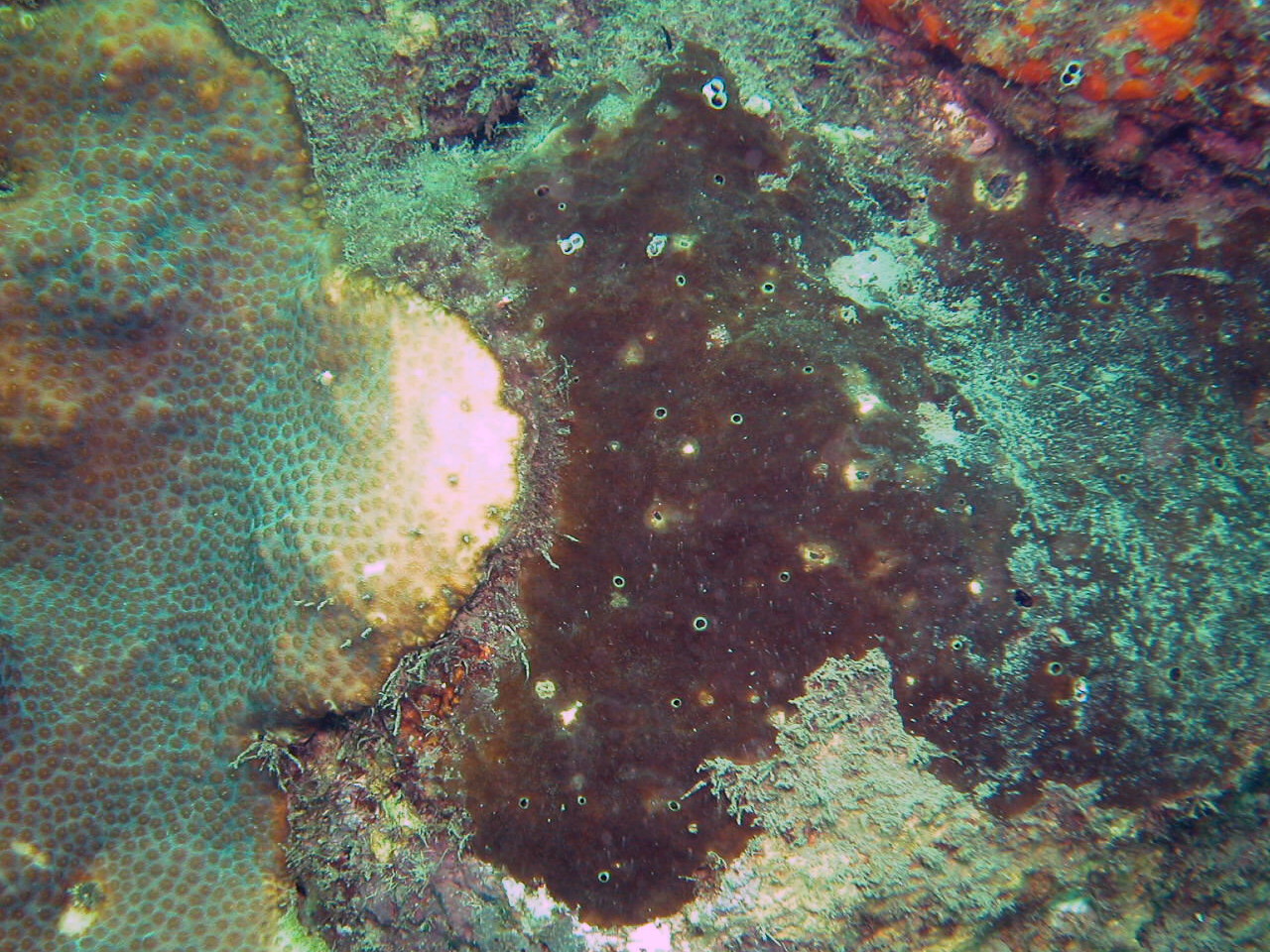
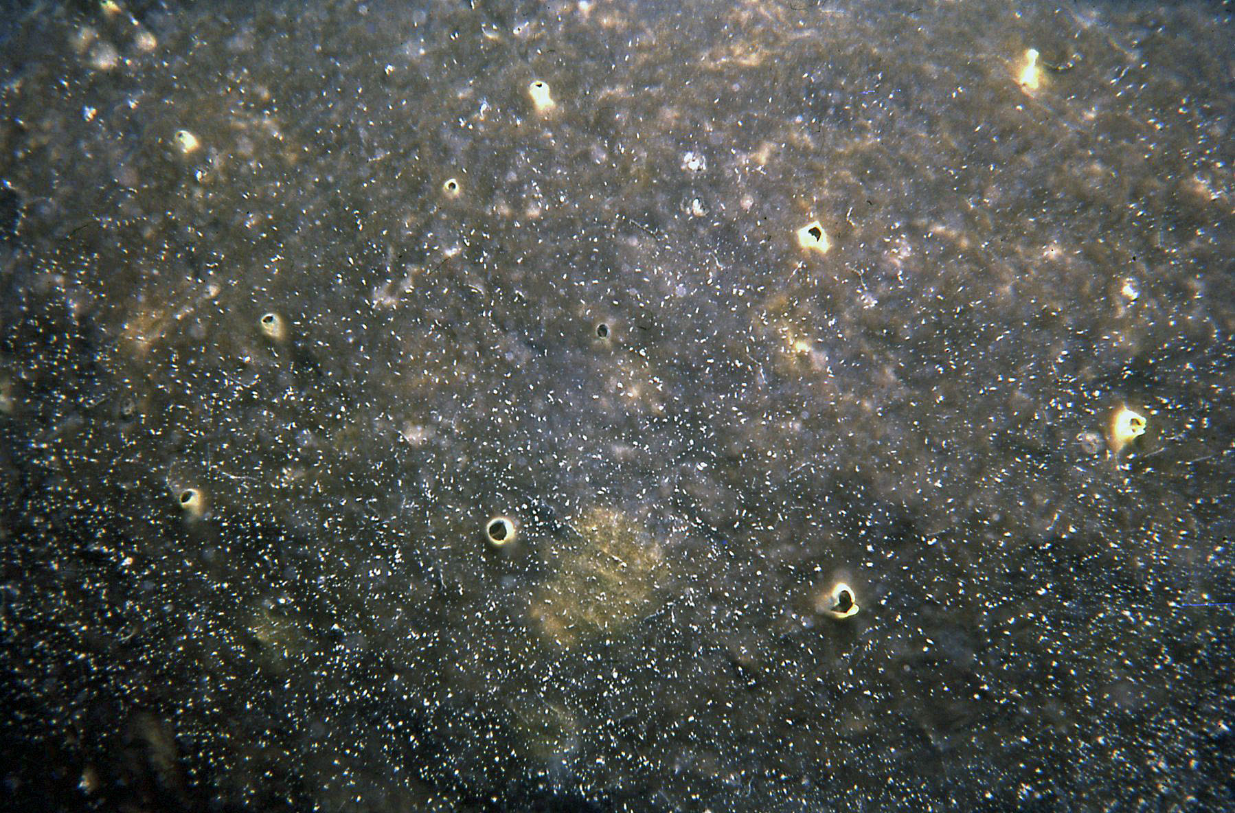
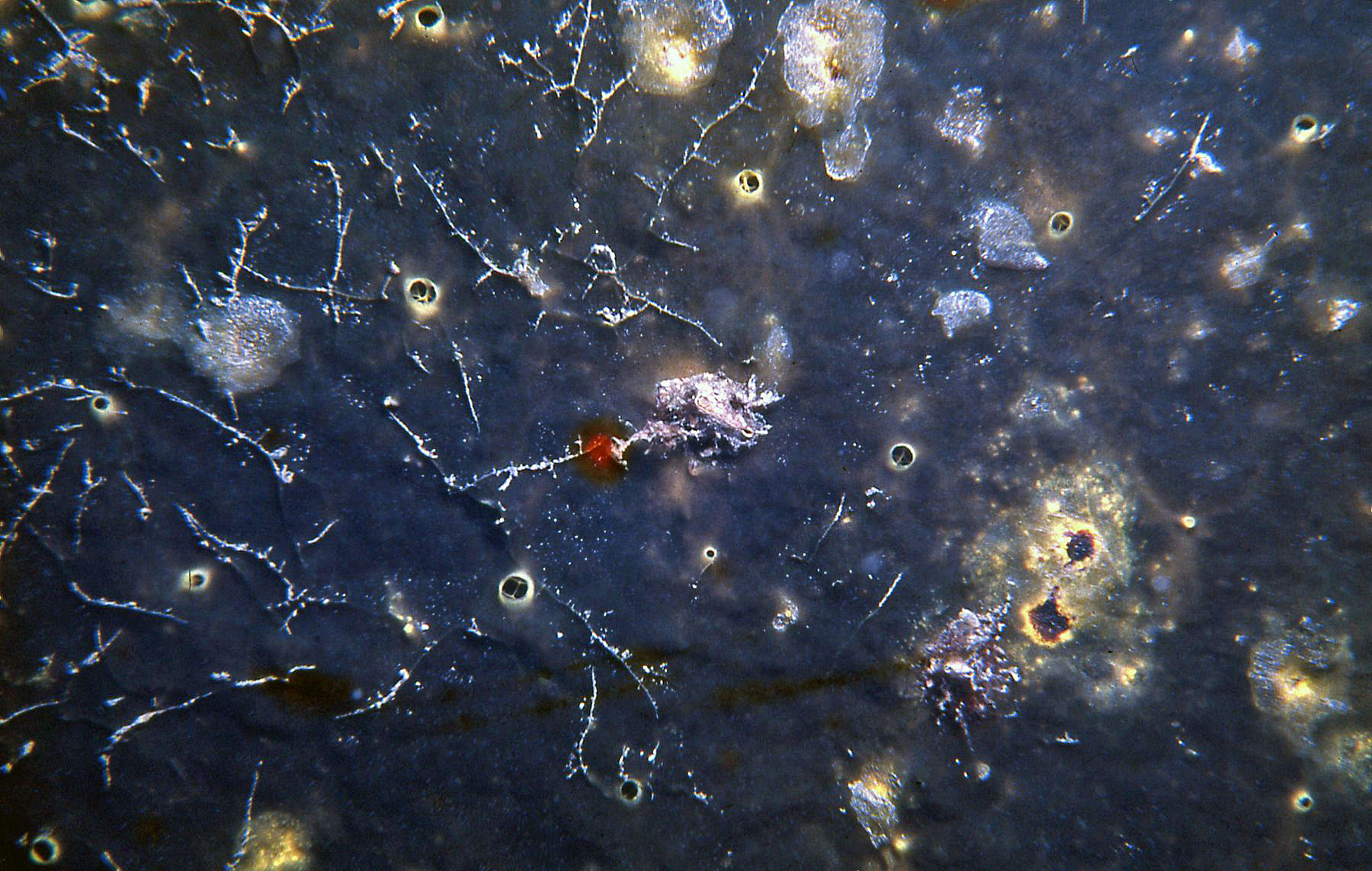
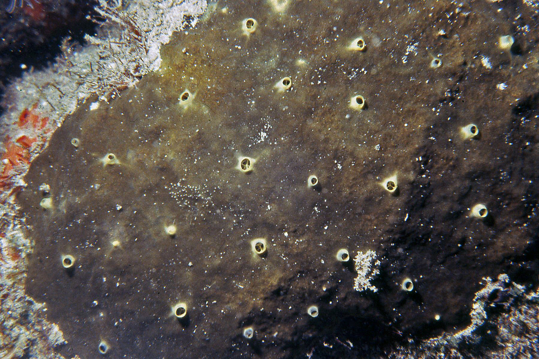
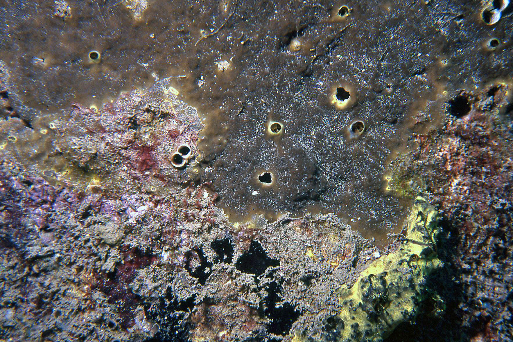
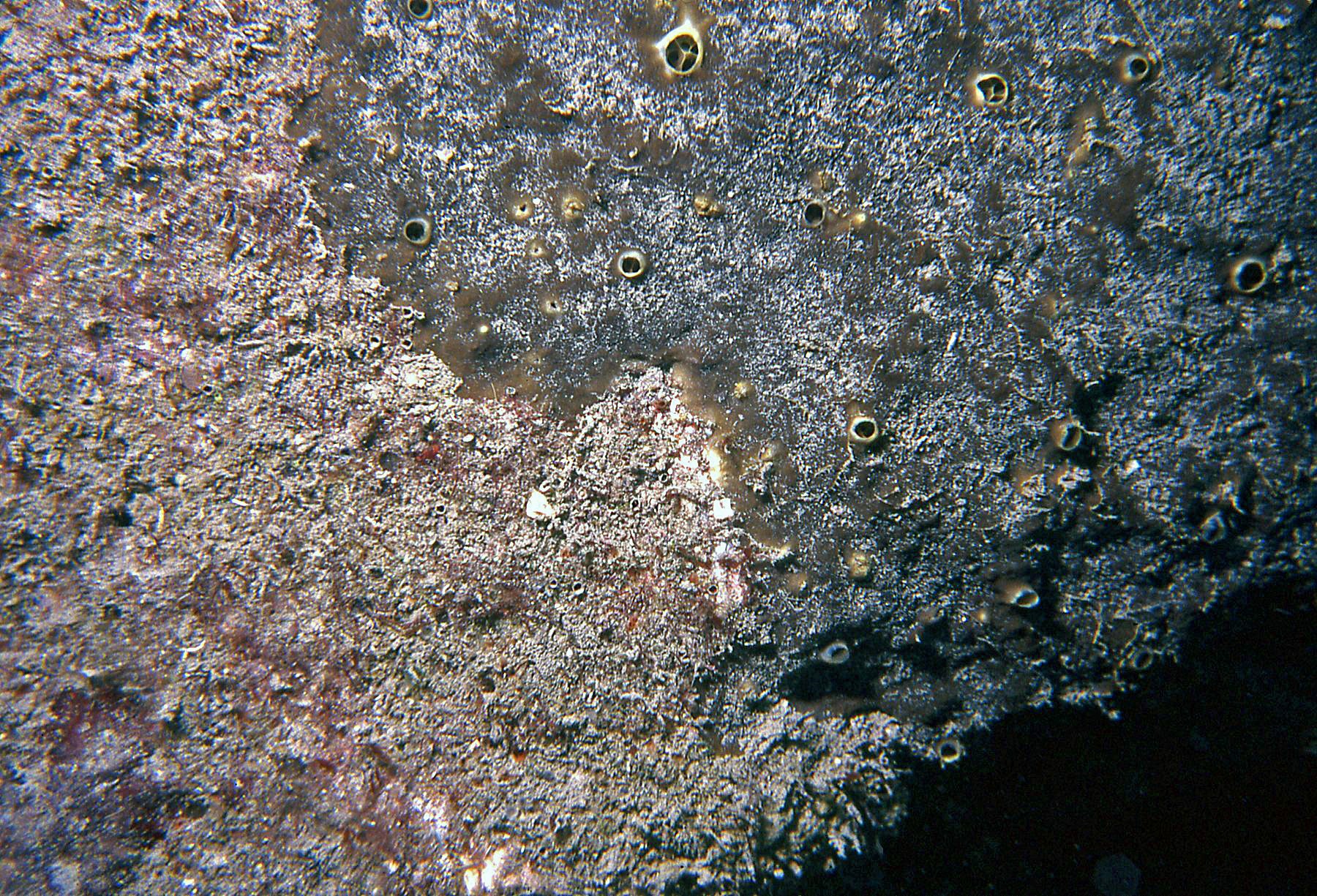
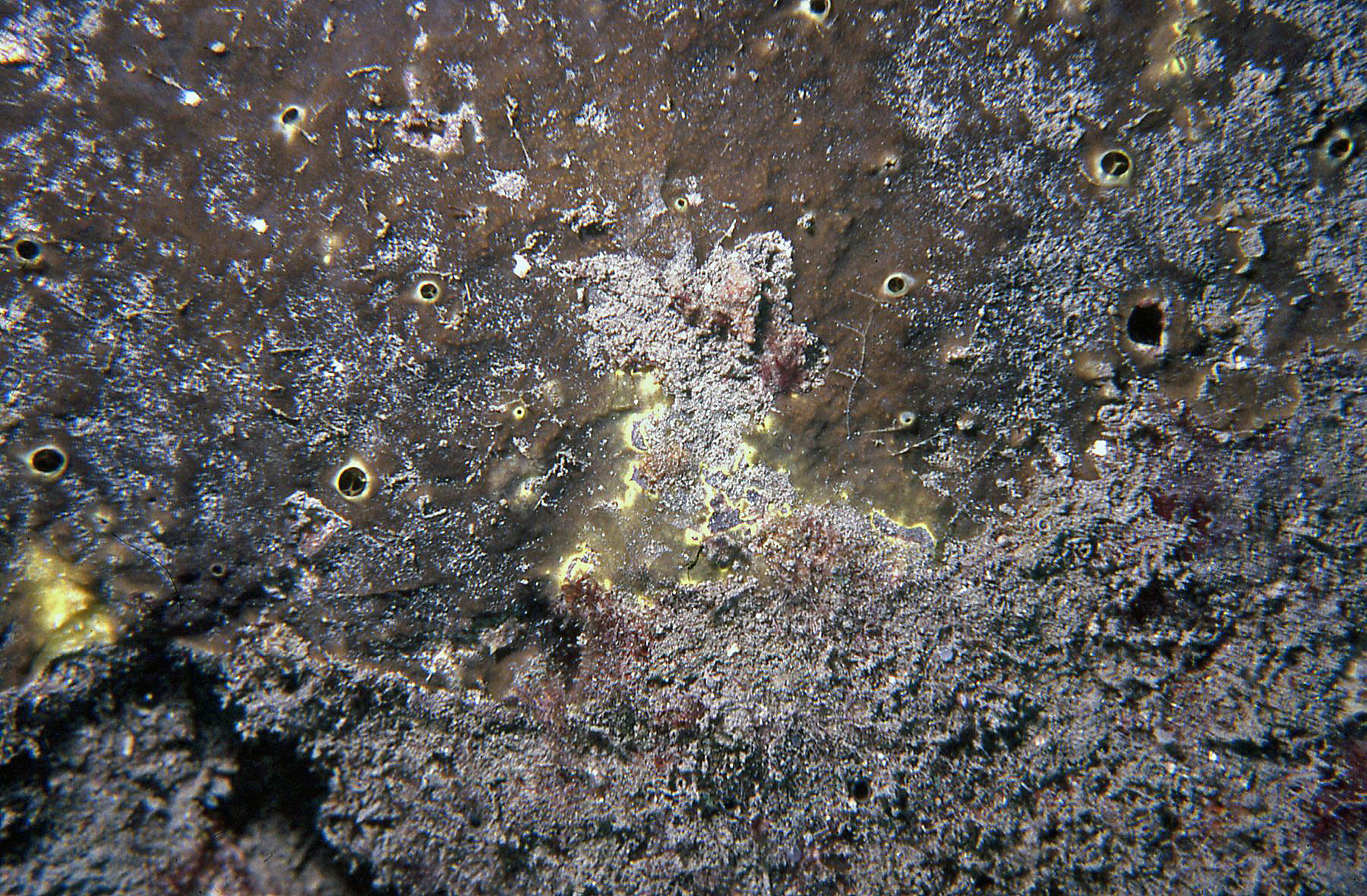
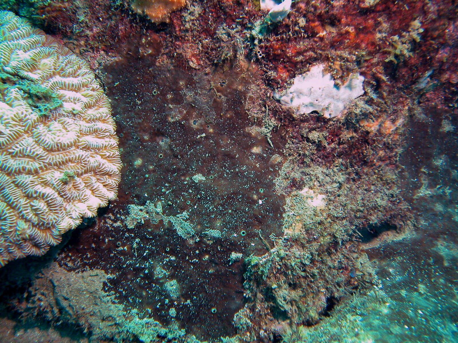
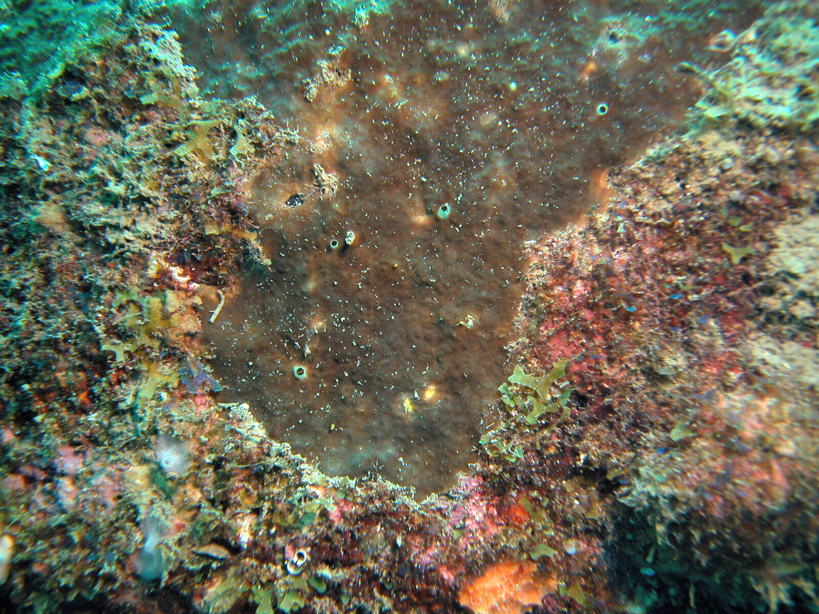
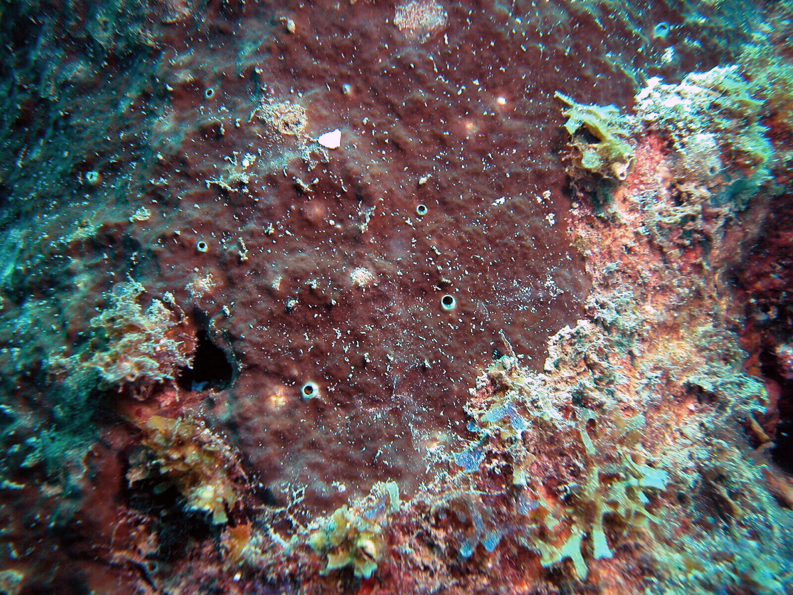
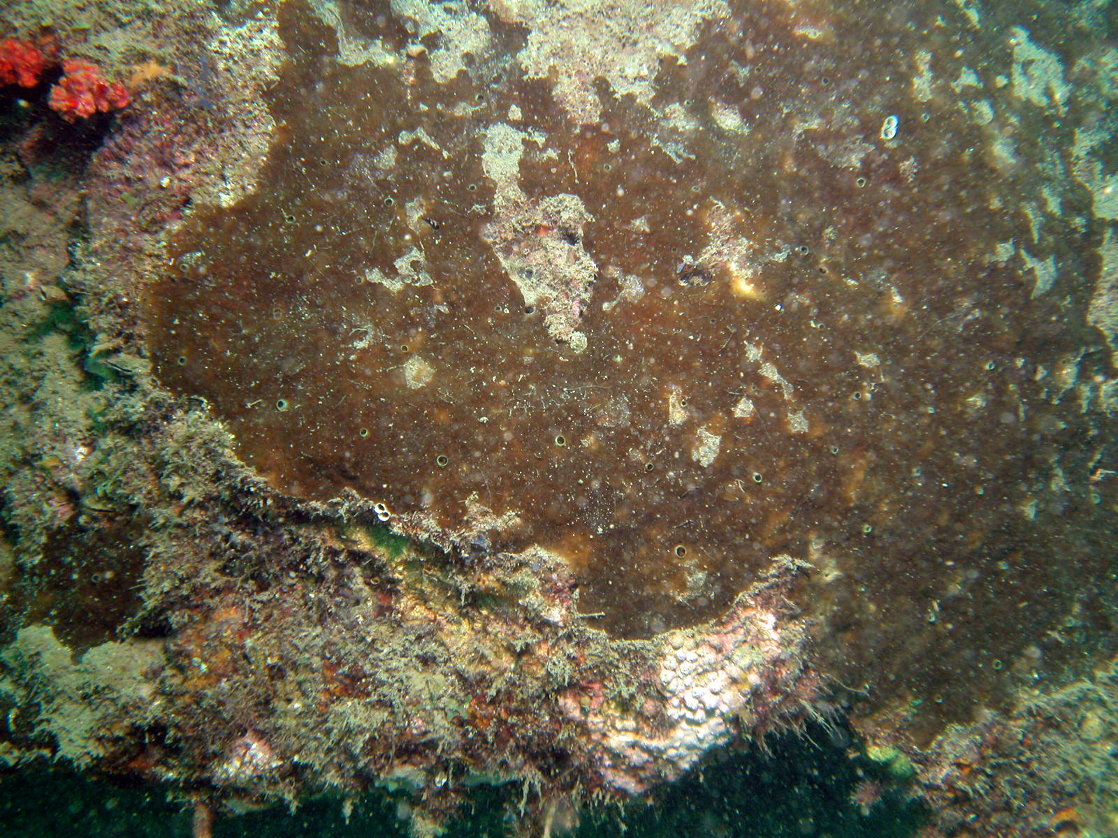
Description: (After Zea & López-Victoria, 2016) Excavating sponge, fully encrusting. It forms a round to irregular epilithic crust, 1-2 mm thick, and from a few cm to about 50-80 cm in maximum diameter. Oscules conspicuous, scattered, 1-8 mm in diameter, with a 1-2 mm tall membranous collar. Surface color amber brown to gray brown, sometimes greenish; oscular collars bright sulfur yellow. Consistency of the ectosome leathery, crumbly for the choanosomal tissue. Surface as even, or as rugose as the underlying substratum; smooth, rather velvety, shiny when not covered by sediment-ladden mucus. Sponge penetrates and excavates about 1-3 cm underneath the surface. Excavating tissue ochre yellow, fills calicular spaces and excavated chambers. Ectosomal skeleton as erect, tree-like plumose tracts up to 8–10 spicules thick, separated at the base 200–300 μm, which ascend and divide 1–2 times up to the surface. Choanosomal skeleton of ill-defined tracts of 2–3 spicules, more often located towards the walls of chambers. Chamber walls creased with concave erosion scars, up to 60 μm wide. Megascleres spicules tylostyles, slender, slightly curved, 280-480 μm long and 5.8-17.5 μm wide; some tylostyles lack a head, but for most the heads are narrow, elongated, more bulged on one side, often misshaped, some subterminal; tips long, acute, most often telescoped. Heads, when present, 6.9-10.3 μm wide and 8.1-15.0 μm long. Microsclere spirasters absent.
Notes: Other Caribbean dark brown fully encrusting excavating sponges of the genus Cliona reaching large sizes are C. caribbaea Carter, 1882, C. tenuis Zea & Weil, 2003 and C. varians (Duchassaing & Michelotti, 1864), all pictured here. C. caribbaea and C. tenuis always possess spiraster microscleres (lacking in C. acephala) and the heads of the tylostyles are always clearly delineated and rarely deformed, usually drop-shaped to rounded, in contrast to the narrow to deformed or absent heads in C. acephala. Tylostyle dimensions of C. acephala (280–480 x 5.8–17.5 μm) overlap with those of C. caribbaea (271–418 x 4.7–15.2 μm) but are larger than in C. tenuis (199–380 μm x 3.3–14.3 μm) (Zea & Weil 2003). While C. caribbaea can occur in papillate morphology, its encrusting form is like that of C. acephala and develops a similar thickness of the epilithic crust and has a similar oscular diameter, but the tylostyle shape is very different between the two species (Zea & Weil 2003). Although reef-dwelling specimens of C. varians are usually encrusting (also called forma incrustans Wiedenmayer, 1977) and tan to yellow brown, the few that are dark brown can potentially be confused in the field with C. acephala. C. varians is distinguished by a greater thickness of the epilithic crust (up to 3–6 mm), much larger oscules (up to 1 cm in diameter) and its characteristic C-shaped spirasters (also called anthosigmas). Phylogenetic analysis of nuclear ribosomal internal transcribed spacer 2 (ITS2) DNA sequences carried out by Escobar et al. (2012) showed C. acephala as genetically distinct from the other Caribbean congeners, and more closely related to Indo-Pacific C. orientalis Thiele, 1900.
Author Reference: Zea & López-Victoria, 2016
Link: World Porifera Database
Tissue and Spicule Images
Images
acephala
- Location: Colombia-Santa Marta
- Photographer: Sven Zea
- Location: Colombia-Santa Marta
- Photographer: Sven Zea
- Location: Colombia-Santa Marta
- Photographer: Sven Zea
- Location: Colombia-Santa Marta
- Photographer: Sven Zea
- Location: Colombia-Santa Marta
- Photographer: Sven Zea
- Location: Colombia-Santa Marta
- Photographer: Sven Zea
- Location: Colombia-Santa Marta
- Photographer: Sven Zea
- Location: Colombia-Santa Marta
- Photographer: Sven Zea
- Location: Colombia-Santa Marta
- Photographer: Sven Zea
- Location: Colombia-Santa Marta
- Photographer: Sven Zea
- Location: Colombia-Santa Marta
- Photographer: Sven Zea
- Location: Colombia-Santa Marta
- Photographer: Sven Zea
- Location: Colombia-Santa Marta
- Photographer: Sven Zea
- Location: Colombia-Santa Marta
- Photographer: Sven Zea