Observed Characteristics:
Color:
- red
- orange
Morphology:
- branching
- bushy
- encrusting
- fan
- lobate
- massive
Consistency:
- crumbly
- tough
Locations:
- Bahamas
- Colombia
- United States
Species Description and Notes
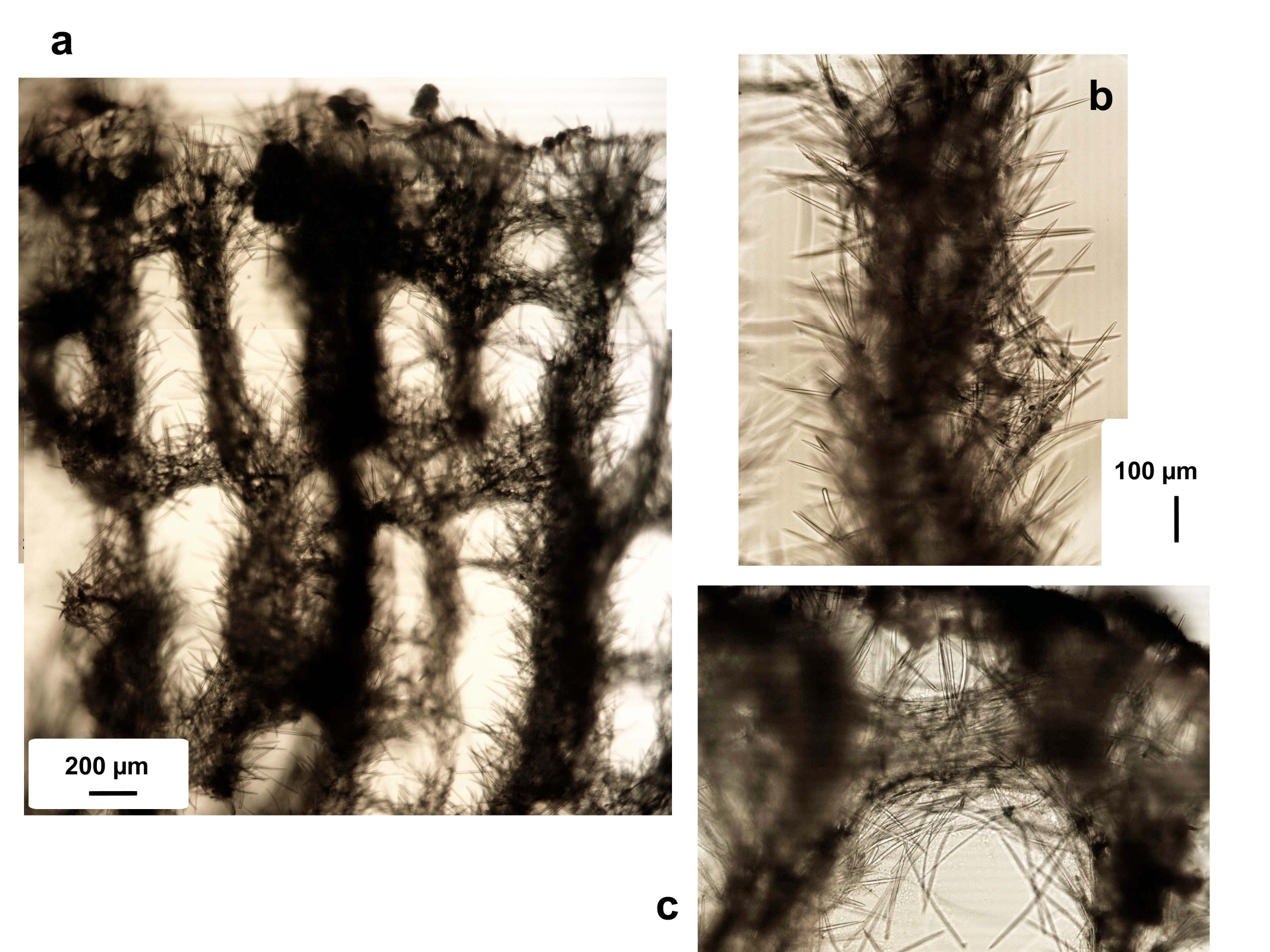
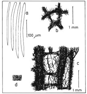
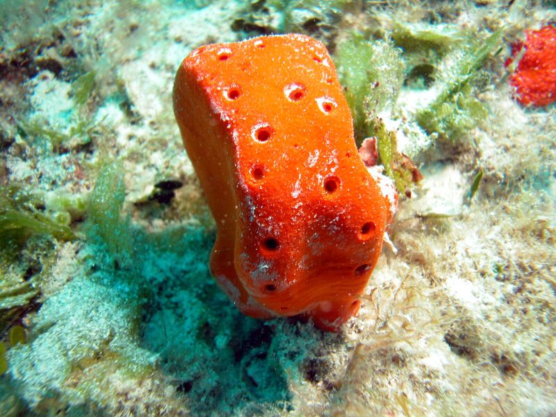
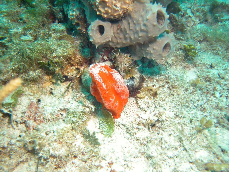
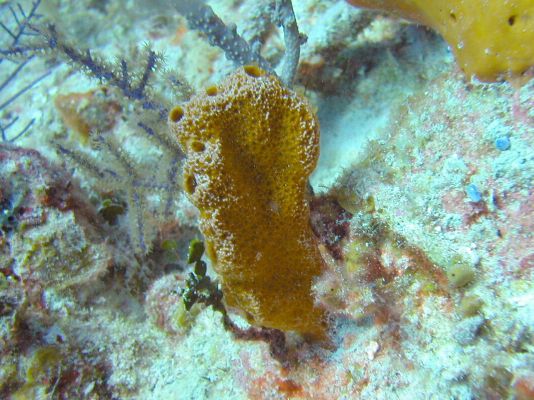
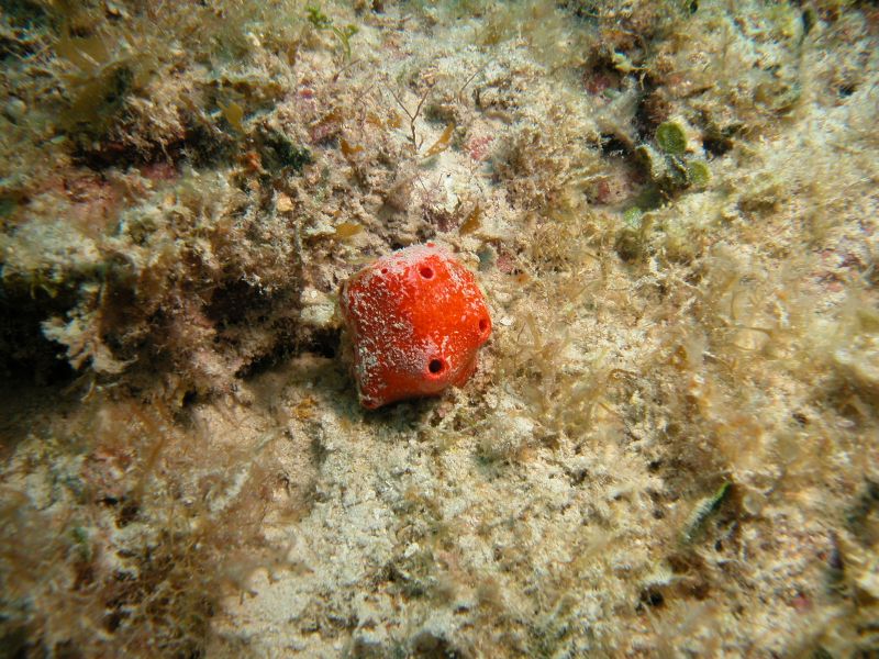
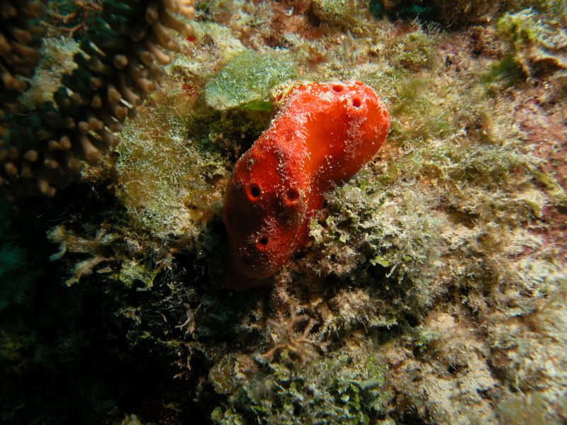
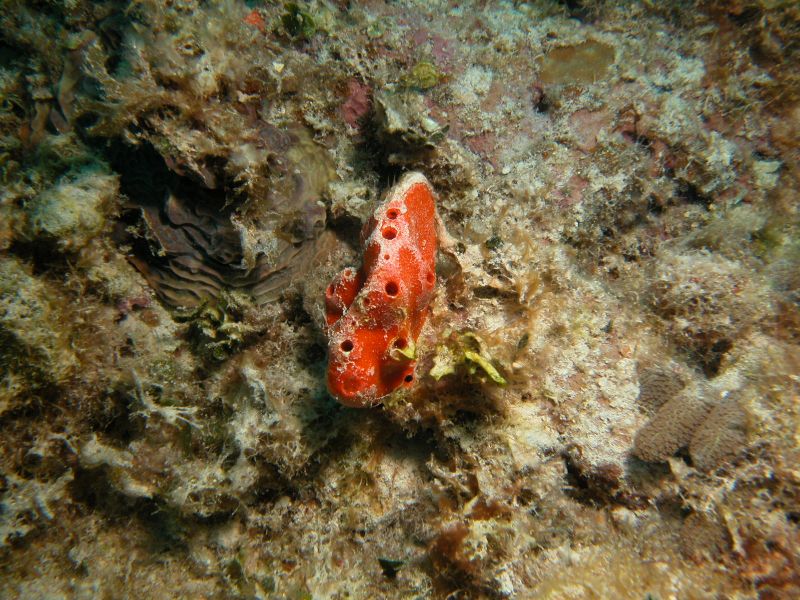
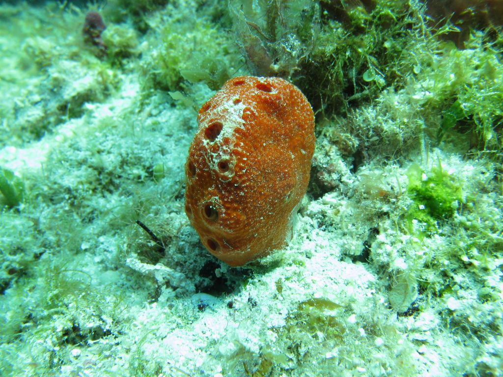
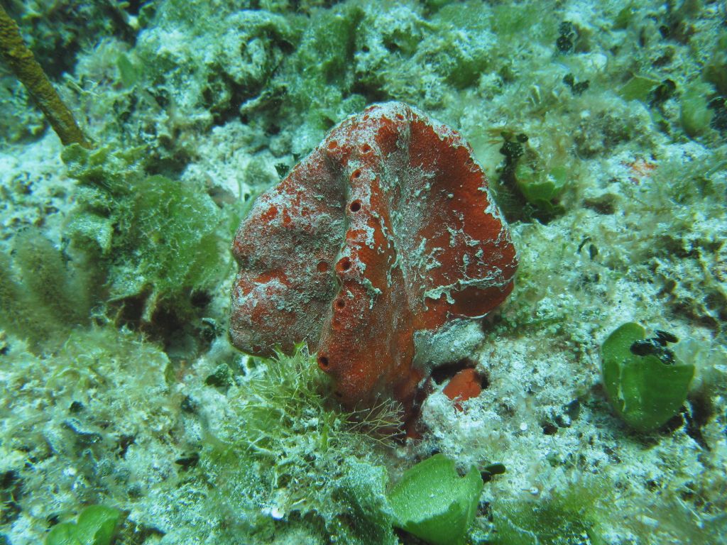
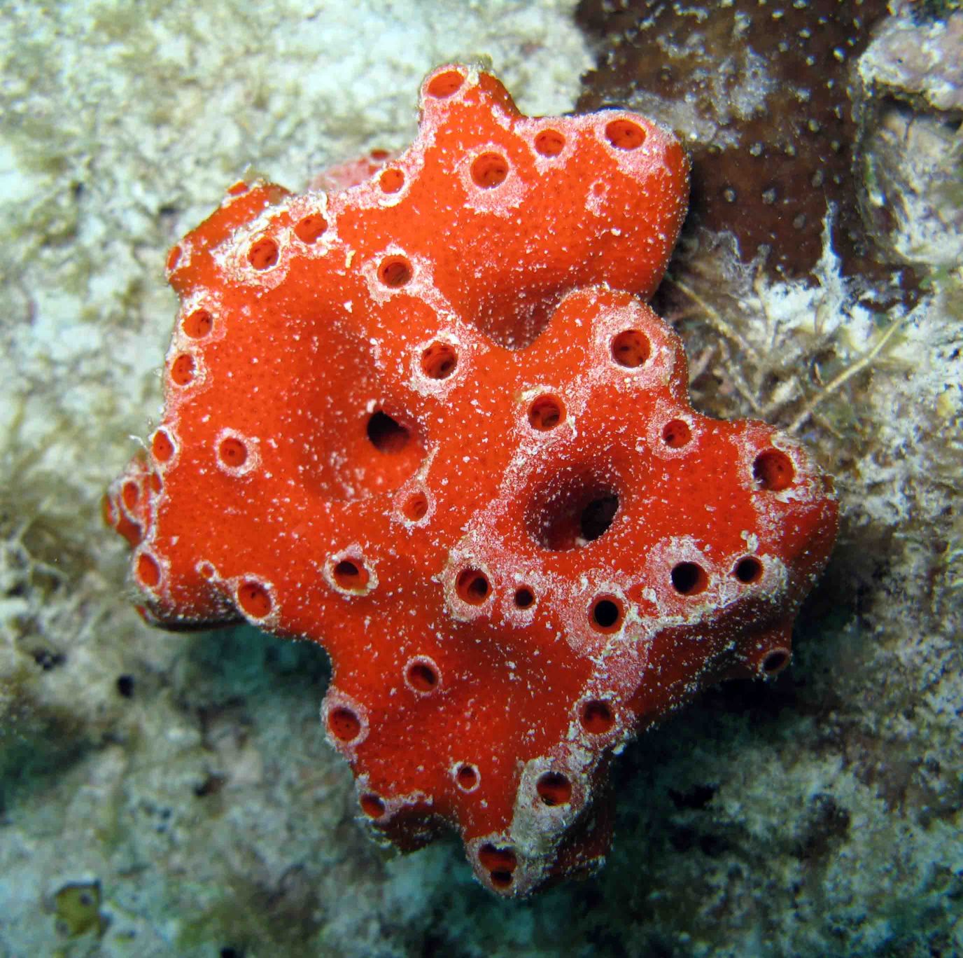
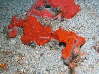
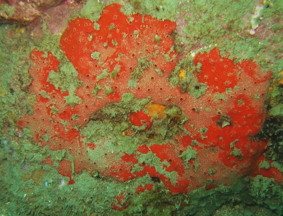
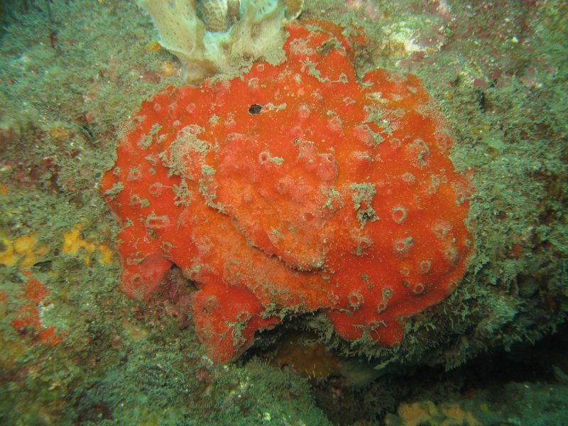
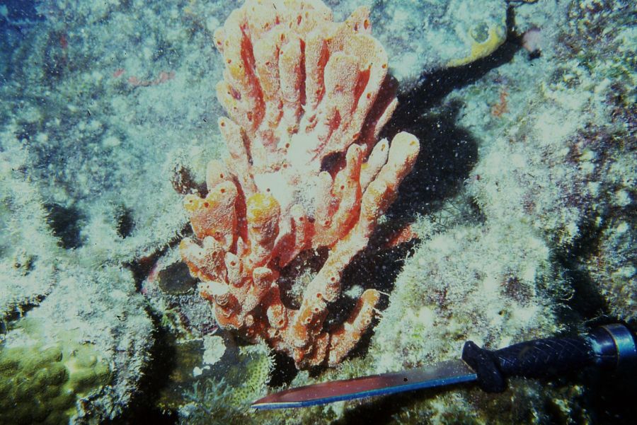
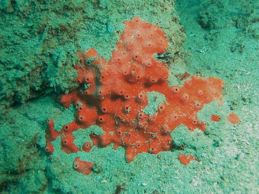
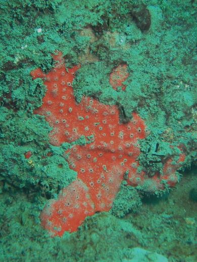
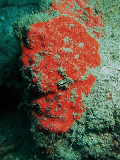
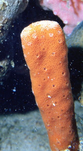
Description: Small masses to erect clubs or thick lamellae; large specimens can form a bouquet of branches thickened at the ends. Size from a few cm wide and tall, to 25-30 cm tall or more when branching. At Santa Marta, Colombia, it is always thickly encrusting, 2-3 cm thick, up to 20-25 cm in diameter. Surface is usually smooth, microspined, although it may be more strongly spined in elevated portions; oscules flush to the surface or slightly elevated, scattered, or aligned on the upper ridges where these occur. External color is bright red scarlet to orange, internal color orange. Consistency firm, somewhat compressible and flexible, but breaks or crumbles when forced. When handled, specimens release abundant mucus. Skeleton as a vertically elongated prismatic reticulation of thick ascending and diverging columnar multispicular tracts, 100-500 µm wide, separated up to 300 µm, interconnected every 175-1,225 µm by transversal spicular tracts, with less packed with spicules, 50-350 µm thick. All tracts are strongly echinated by spicules. At the surface, ascending tracts end in spicule brushes that support the organic pinacoderm. Spicules are: (1) curved styles with smooth heads and acute to slightly telescopic ends, 200-300 µm long by 4.5-13 µm wide; (2) curved, hastate oxea, with short ends, usually slightly asymmetric, slightly longer but thinner than the styles, 260-340 µm long by 7,5-12 µm thick.
Notes: This is a shallow to deep reef species, living usually exposed. Previously known as Pseudaxinella lunaecharta (Ridley & Dendy, 1886) (e.g., Wiedenmayer, 1977; Zea, 1987), it was synonymized to Pseudaxinella reticulata (Ridley & Dendy, 1886) from Brazil by Alvarez et al. (1998), which was then transferred to genus Dragmacidon by Alvarez & Hooper (2002). However, Zea & Pulido (2016) found that there are two distinct morphotypes that fall under the current definition of D. reticulatum, one corresponding to the actual D. reticulatum, also pictured here, and the other into D. lunaecharta sensu Wiedenmayer (1977). These authors erected a new name for the latter, vis. D. alvarezae, as the former name corresponds to a valid and distinct Eastern Atlantic species. Dragmacidon reticulatum is clearly distinct from D. alvarezae in overall shape and spicule characteristics. They coexist in some areas (Stirrups Cays, Bahamas, Santa Marta, Colombia). D. alvarezae has a smoother surface and often grows erect, while D. reticulatum has a more elaborated surface, always with thick spines, and is predominantly thickly encrusting and slightly tougher. In coexisting specimens, typically D. alvarezae has smaller spicules, with the oxea larger than the styles, while D. reticulatum has larger spicules and some of the styles as long as the oxea. The skeleton of D. reticulatum is made of thicker ascending spicule tracts, more widely separated, and projecting on the surface as spines. [Note of caution: in the 3rd. edition of this guide we switched the names for the two morphotypes of Dragmacidon: D. reticulatum for D. “lunaecharta” and D. explicatum (Wiedenmayer, 1977) for the actual D. reticulatum (according to Alvarez et al., 1998, and to the examination of types by Zea & Pulido (2016), D. explicatum is a junior synonym of D. reticulatum.)]
Author Reference: Zea & Pulido, 2016
Link: World Porifera Database
Tissue and Spicule Images

Images
alvarezae
- Location: Stirrups Cays, N Berry Islands, Bahamas
- Photographer: Sven Zea
- Location: Stirrups Cays, N Berry Islands, Bahamas
- Photographer: Sven Zea
- Location: Little San Salvador, Bahamas
- Photographer: Sven Zea
- Location: Stirrups Cays, N Berry Islands, Bahamas
- Photographer: Sven Zea
- Location: Stirrups Cays, N Berry Islands, Bahamas
- Photographer: Sven Zea
- Location: Stirrups Cays, N Berry Islands, Bahamas
- Photographer: Sven Zea
- Location: Stirrups Cays, N Berry Islands, Baham
- Photographer: Sven Zea
- Location: Stirrups Cays, N Berry Islands, Bahamas
- Photographer: Sven Zea
- Location: Florida Keys, United States
- Photographer: Joseph Pawlik
- Location: Islas del Rosario, Colombia
- Photographer: Sven Zea
- Location: Santa Marta, Colombia
- Photographer: Sven Zea
- Location: Santa Marta, Colombia
- Photographer: Sven Zea
- Location: Santa Marta, Colombia
- Photographer: Sven Zea
- Location: Santa Marta, Colombia
- Photographer: Sven Zea
- Location: Islas del Rosario, Colombia
- Photographer: Sven Zea
- Location: Santa Marta, Colombia
- Photographer: Sven Zea
- Location: Santa Marta, Colombia
- Photographer: Sven Zea
- Location: Santa Marta, Colombia
- Photographer: Sven Zea
- Location: Islas del Rosario, Colombi
- Photographer: Sven Zea